Review
. 2020 Dec 11;10(12):2377.
doi: 10.3390/ani10122377.

Pathology of Coronavirus Infections: A Review of Lesions in Animals in the One-Health Perspective

Affiliations
Review

Pathology of Coronavirus Infections: A Review of Lesions in Animals in the One-Health Perspective

Valentina Zappulli et al. Animals (Basel). .

Abstract

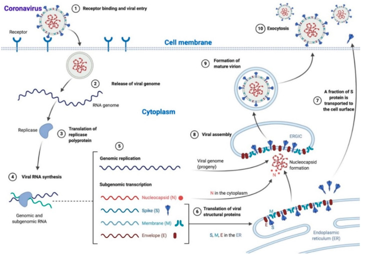
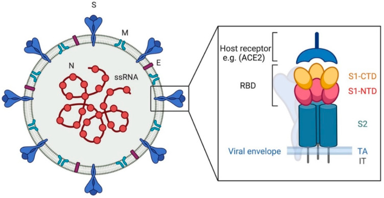
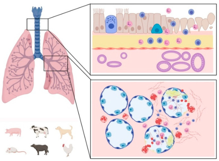
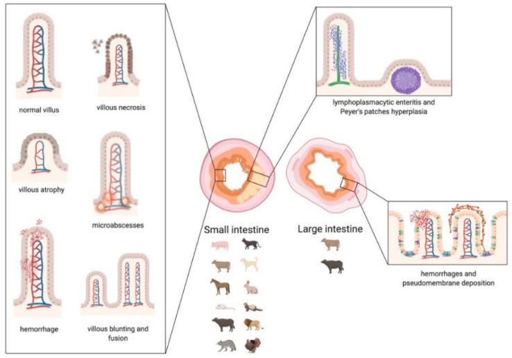
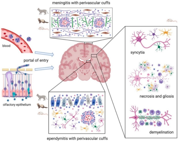
Coronaviruses (CoVs) are worldwide distributed RNA-viruses affecting several species, including humans, and causing a broad spectrum of diseases. Historically, they have not been considered a severe threat to public health until two outbreaks of COVs-related atypical human pneumonia derived from animal hosts appeared in 2002 and in 2012. The concern related to CoVs infection dramatically rose after the COVID-19 global outbreak, for which a spill-over from wild animals is also most likely. In light of this CoV zoonotic risk, and their ability to adapt to new species and dramatically spread, it appears pivotal to understand the pathophysiology and mechanisms of tissue injury of known CoVs within the "One-Health" concept. This review specifically describes all CoVs diseases in animals, schematically representing the tissue damage and summarizing the major lesions in an attempt to compare and put them in relation, also with human infections. Some information on pathogenesis and genetic diversity is also included. Investigating the lesions and distribution of CoVs can be crucial to understand and monitor the evolution of these viruses as well as of other pathogens and to further deepen the pathogenesis and transmission of this disease to help public health preventive measures and therapies.

Keywords: One Health; coronavirus; pathology; veterinary medicine.


Conflict of interest statement

The authors declare no conflict of interest.

Figures

Scheme 1
Scheme 1
Coronavirus life cycle. Most Coronaviruses enter the cell after S protein–receptor interaction (1), the viral genome is released in the cytoplasm (2) and translated into the replicase polyprotein (3) to synthetize viral RNA (4). Genomic (progeny) and subgenomic RNAs are produced (5) and the latter are translated to structural and accessory proteins that can be inserted in the endoplasmic reticulum (ER; 6) and moved to the endoplasmic reticulum-Golgi intermediate compartment (ERGIC), while a fraction of S protein is transported directly to the cell surface where it mediates cell–cell fusion (7). In the ERGIC, the viral assembly and the encapsidation of genomic RNA by N protein take place, leading to nucleocapsid formation (8). Mature virions are then transported in smooth-walled vesicles (9) and released via exocytosis (10). N: nucleocapsid, S: spike, M: membrane, E: envelope. The scheme has been created with BioRender.com.
Scheme 2
Scheme 2
Coronavirus, viral particle. The image shows, on the left, a graphic representation of a viral particle. The black box shows the structure of the spike protein. N: nucleocapsid, S: spike, M: membrane, E: envelope. RBD: receptor binding domain, S1-CTD: subunit 1 C-terminal domain, S1-NTD: subunit 1 N-terminal domain, S2: subunit 2, TA: transmembrane anchor, IT: intracellular tail. The scheme has been created with BioRender.com.
Scheme 3
Scheme 3
Graphic representation of the main respiratory lesions associated with Coronavirus infections in animals. Affected upper (top box) and lower (bottom box) respiratory tract: the healthy tissue is represented on the left side of the boxes and the lesions on the right side. The species mainly affected by respiratory lesions are represented on the lower left corner. Lesions of the upper airways are mainly characterized by mucosal inflammation consisting of neutrophilic (or heterophilic) and lymphocytic infiltrate, sloughing of epithelial cells, loss of cilia, epithelial cell hyperplasia, and hypertrophy of mucosal glands. Lesions in the lungs are mainly characterized by hemorrhages with neutrophilic (or heterophilic) and lymphocytic infiltrate, severe edema, epithelial cell detachment, and fibrinous exudate. The scheme has been created with BioRender.com.
Scheme 4
Scheme 4
Graphic representation of the main lesions detected in small and large intestine associated with Coronavirus infections in animals. Small intestine: in the left box, the morphologic changes of the villi are represented. In the upper right box, the distribution of the infiltrate, affecting the lamina propria and the submucosa, is represented. Large intestine: in the lower right box, the lesions affecting the mucosa of the colon are represented. The main categories of affected species that suffer damage mainly to the small or large intestine are indicated below the corresponding image. The scheme has been created with BioRender.com.
Scheme 5
Scheme 5
Graphic representation of the main brain lesions associated with Coronavirus infections in animals. Typically, the virus disseminates to the brain either via the blood within monocytes or along the olfactory nerve through the olfactory epithelium (left of the image). Main lesions affecting cats, ferrets, and mice are represented by meningitis and ependymitis with perivascular cuffs. Lesions in mice are also characterized by syncytia of neurons, astrocytes, microglia, and oligodendrocytes. Necrosis and gliosis are common features in laboratory animals. Chronic infections in mice can result in demyelination. The scheme has been created with BioRender.com.
Figure 1
Figure 1
Feline infection peritonitis (FIP), liver (L), cat. Hepatic parenchyma is effaced by a multifocal to coalescent mixed inflammatory infiltrate (arrows) with central necrosis (asterisks). Hematoxylin eosin stain. Inset: Immunohistochemistry shows intense brown cytoplasmic immunolabelling for feline CoV antigen within macrophages.

References

    1. Wang C., Horby P.W., Hayden F.G., Gao G.F. A novel coronavirus outbreak of global health concern. Lancet. 2020;395:470–473. doi: 10.1016/S0140-6736(20)30185-9. - DOI - PMC - PubMed
    1. Wu D., Wu T., Liu Q., Yang Z. The SARS-CoV-2 outbreak: What we know. Int. J. Infect. Dis. 2020;94:44–48. doi: 10.1016/j.ijid.2020.03.004. - DOI - PMC - PubMed
    1. Cunningham A.A., Daszak P., Wood J.L.N. One Health, emerging infectious diseases and wildlife: Two decades of progress? Philos. Trans. R. Soc. B Biol. Sci. 2017;372:20160167. doi: 10.1098/rstb.2016.0167. - DOI - PMC - PubMed
    1. Kreuder Johnson C., Hitchens P.L., Smiley Evans T., Goldstein T., Thomas K., Clements A., Joly D.O., Wolfe N.D., Daszak P., Karesh W.B., et al. Spillover and pandemic properties of zoonotic viruses with high host plasticity. Sci. Rep. 2015;5:14830. doi: 10.1038/srep14830. - DOI - PMC - PubMed
    1. Sleeman J.M., DeLiberto T., Nguyen N. Optimization of human, animal, and environmental health by using the One Health approach. J. Vet. Sci. 2017;18:263. doi: 10.4142/jvs.2017.18.S1.263. - DOI - PMC - PubMed

LinkOut - more resources