Review
. 2020 Dec 12;21(24):9458.
doi: 10.3390/ijms21249458.

Supramolecular Architectures of Nucleic Acid/Peptide Hybrids

Affiliations
Review

Supramolecular Architectures of Nucleic Acid/Peptide Hybrids

Sayuri L Higashi et al. Int J Mol Sci. .

Abstract

Supramolecular architectures that are built artificially from biomolecules, such as nucleic acids or peptides, with structural hierarchical orders ranging from the molecular to nano-scales have attracted increased attention in molecular science research fields. The engineering of nanostructures with such biomolecule-based supramolecular architectures could offer an opportunity for the development of biocompatible supramolecular (nano)materials. In this review, we highlighted a variety of supramolecular architectures that were assembled from both nucleic acids and peptides through the non-covalent interactions between them or the covalently conjugated molecular hybrids between them.

Keywords: molecular hybrid; nanostructures; nucleic acids; peptides; self-assembly; supramolecular architectures.


Conflict of interest statement

The authors declare no conflict of interest. The funders had no role in the design of the study; in the collection, analyses, or interpretation of data; in the writing of the manuscript, or in the decision to publish the results.

Figures

Figure 1
Figure 1
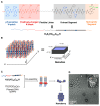
(A) DNA and (B) peptide nanotechnologies. Representative supramolecular architectures (nanostructures) and their typical component structural motifs are shown.
Figure 2
Figure 2
(A) Chemical structure of the self-assembling peptide derivatives containing K3, a self-assembling β-sheet domain, and a hydrophilic segment at the C-terminus for controlled water dispersibility. (B) Schematic illustration of the self-assembly of the peptide derivatives to form the nanoribbons. (C) (a) Schematic illustration of the co-assembly of the peptide derivatives with plasmid DNA (pDNA) to form a variety of hybrid nanostructures. (b) Cross-sectional view of the nanococoons. Adapted from [58]. Copyright 2014 American Chemical Society. Adapted from [59]. Copyright 2017 John Wiley and Sons Publisher.
Figure 3
Figure 3
(A) Chemical structure of the self-assembling peptide derivative, H4K5HCBzlCBzlH. (B) Schematic illustration of a nanodisc that was constructed via the self-assembly of H4K5HCBzlCBzlH. (C) Schematic illustration and (D) representative TEM image (magnified in inset) of the co-assembly between H4K5HCBzlCBzlH with pDNA to form the nanoberries. Adapted from [60]. Copyright 2020 John Wiley and Sons Publisher.
Figure 4
Figure 4
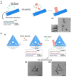
(A) (i) Schematic illustration of the formation of a semi-artificial tobacco mosaic virus (TMV) nanostructure at a designated position of the DNA origami nanotube and (ii) representative TEM images of the in situ assembled protein nanotubes on DNA origami nanotubes with one docking site. (B) (i) Schematic illustration of the spatiotemporally controlled TMV protein assembly on the surface of a DNA origami triangle, (ii,iii) representative TEM images of each state illustrated in (i). Scale bar: 50 nm. Adapted from [64,65]. Copyright 2018 and 2020 American Chemical Society.
Figure 5
Figure 5
(A) Design of SP-CC-PEG to form mushroom-shaped nanostructures. (B) Schematic illustration of the formation of supramolecular rod-like nanostructures through the complexation of SP-CC-PEG5000 with dsDNAs. Adapted from [66]. Copyright 2013 American Chemical Society.
Figure 6
Figure 6
(A) Sequences of the three collagen-like triple helix-forming peptides (CP+, CP++, and sCP++). (B) Schematic illustration of the co-assembly of the two-layered DNA origami nanosheets (DNA TL nanosheets) and the collagen-mimetic peptides (CP++ and sCP++) to obtain the one-dimensional peptide/DNA hybrid nanowires. (C) Representative TEM images of assembled with DNA TL nanosheets and CP++. Scale bar: 50 nm. Adapted from [68]. Copyright 2017 American Chemical Society.
Figure 7
Figure 7
(A) Sequences of the coiled-coil heterodimers that formed the peptides (CC-Di-EK and CC-Di-KE) that were conjugated to the oligonucleotides (α, β). (B) Schematic illustration and (C) a representative TEM image of the connection of two distinct one-dimensional DNA origami nanostructures (Origamis A and B) by the specific formation of peptide coiled-coil heterodimers. (D) Sequences of the coiled-coil heterodimers that formed the peptides (EI and KI) that were conjugated to the oligonucleotides (DNA A and B). (E) Schematic illustration and (F) representative AFM images to obtain long one-dimensional arrays of DNA origami cuboid nanostructures. Scale bar: 50 nm for C and 1 μm (250 nm (inset)) for F. Adapted from [69,70]. Copyright 2019 and 2020 American Chemical Society.
Figure 8
Figure 8
(A) Schematic illustration and (B) a representative AFM image of amyloid fibril-filled DNA origami nanotubes that were organized onto two-dimensional DNA origami platforms. Scale bars: 250 nm. The colour scale bar indicates height. Adapted from [75]. Copyright 2014 Springer Nature.
Figure 9
Figure 9
(A) Schematic illustration of the orthogonal self-assembly of semi-artificial glycopeptides (right panel) and ssDNAs (left panel) to produce hybrid soft materials that comprised supramolecular nanostructures of glycopeptide and DNA microspheres. (B) Representative confocal laser scanning microscopy (CLSM) images showing the orthogonal coexistence of DNA microsphere and the supramolecular nanostructures of (a) Z-FF-Mal, (b) Z-FF-Cel, and (c) Z-FF-Lac. Scale bar: 5 µm for a and 10 µm for b,c. Adapted from [76]. Copyright 2019 John Wiley and Sons Publisher.
Figure 10
Figure 10
(A) Chemical structure of ssDNA12-FF. (B) Plausible supramolecular architectures of the hollow vesicular structures that were obtained through the self-assembly of ssDNA12-FF. (C) Chemical structure of ssDNA12-WW. (D) Plausible, concentration-dependent supramolecular architectures of ssDNA12-WW obtained by its self-assembly. Adapted from [91,92]. Copyright 2012 and 2014 Royal Chemical Society.
Figure 11
Figure 11
(A) Schematic representation of the self-assembly of β-annulus fragment (βAF)_dN20 [N = A (βAF_dA20) or T (βAF_dT20)] to form spherical capsular architectures that displayed the nucleic acids at their exterior surfaces. (B) Schematic representation of the self-assembly of βAF_ssDNA23 to form spherical capsular architectures that encapsulated the nucleic acids. Adapted from ref 88. Copyright 2017 John Wiley and Sons Publisher. Adapted from [95]. Copyright 2019 the Chemical Society of Japan.
Figure 12
Figure 12
Sequences of the two DNA/peptide hybrids, β-suRGD-AS and β-suRGD-S, and the two distinct protocols that controlled their assembly pathway to yield the monodispersed toroidal nanostructures (β-suRGD-AS/β-suRGD-S). Adapted from [104]. Copyright 2016 John Wiley and Sons Publisher.
Figure 13
Figure 13
(A) Chemical structure of Fmoc-FF-ssDNAs. (B) Schematic representation of the self-assembly of Fmoc-FF-ssDNAs to form hierarchical nanostructures through DNA hybridization. (C) Representative TEM images obtained from co-assembly of Fmoc-FF-ssDNA19 and Fmoc-FF-ss(as)DNA19. Scale bar: 200 nm. Adapted from [109]. Copyright 2019 American Chemical Society.
Figure 14
Figure 14
(A) Schematic representation of the formation of nanowires comprising a computationally designed protein/dualENH and a short dsDNA. (B) A representative AFM image obtained after mixing dualENH with the dsDNA (25 bp). Adapted from [112]. Copyright 2014 Springer Nature.
Figure 15
Figure 15
Schematic representation of the formation of linear supramolecular architectures that tethered the proteins by exploiting hybridization chain reaction (HCR) of nucleic acids. Adapted from [116]. Copyright 2018 American Chemical Society.
Figure 16
Figure 16
Three-dimensional cage-like supramolecular architecture obtained by the complexation of a homotrimeric protein containing three ssDNAs with a triangular DNA nanostructure bearing three complementary ssDNAs through DNA hybridization. Adapted from [118]. Copyright 2019 American Chemical Society.
Figure 17
Figure 17
(A) Molecular design of the nucleoproteins (RIDC3-10a and RIDC3-10b). (B) Schematic representation and (C) a representative TEM image of the ordered crystalline architectures obtained through the complexation of RIDC3-10a/10b with Zn2+ under a small window of conditions. Scale bar: 5 µm. Adapted from [119]. Copyright 2018 American Chemical Society.

References

    1. Seeman N.C., Sleiman H.F. DNA nanotechnology. Nat. Rev. Mater. 2018;3:17068. doi: 10.1038/natrevmats.2017.68. - DOI
    1. Madsen M., Gothelf K.V. Chemistries for DNA nanotechnology. Chem. Rev. 2019;119:6384–6458. doi: 10.1021/acs.chemrev.8b00570. - DOI - PubMed
    1. Heuer-Jungemann A., Liedl T. From DNA tiles to functional DNA materials. Trends Chem. 2019;1:799–814. doi: 10.1016/j.trechm.2019.07.006. - DOI
    1. Seeman N.C. DNA in a material world. Nature. 2003;421:427–431. doi: 10.1038/nature01406. - DOI - PubMed
    1. Seeman N.C. Nucleic acid junctions and lattices. J. Theor. Biol. 1982;99:237–247. doi: 10.1016/0022-5193(82)90002-9. - DOI - PubMed

MeSH terms

LinkOut - more resources