Review
. 2020 Dec 12;9(12):2677.
doi: 10.3390/cells9122677.

NOTCH1 Signaling in Head and Neck Squamous Cell Carcinoma

Affiliations
Review

NOTCH1 Signaling in Head and Neck Squamous Cell Carcinoma

Pooja A Shah et al. Cells. .

Abstract

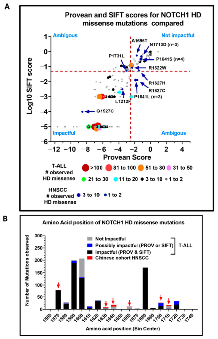
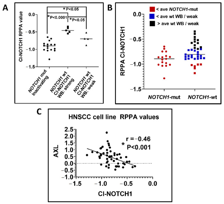
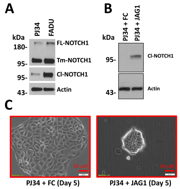
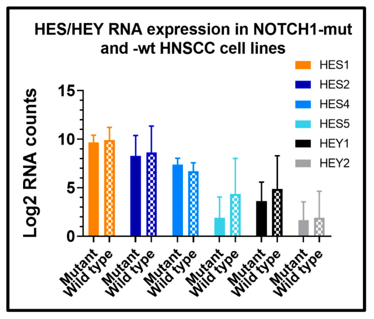
Biomarker-driven targeted therapies are lacking for head and neck squamous cell carcinoma (HNSCC), which is common and lethal. Efforts to develop such therapies are hindered by a genomic landscape dominated by the loss of tumor suppressor function, including NOTCH1 that is frequently mutated in HNSCC. Clearer understanding of NOTCH1 signaling in HNSCCs is crucial to clinically targeting this pathway. Structural characterization of NOTCH1 mutations in HNSCC demonstrates that most are predicted to cause loss of function, in agreement with NOTCH1's role as a tumor suppressor in this cancer. Experimental manipulation of NOTCH1 signaling in HNSCC cell lines harboring either mutant or wild-type NOTCH1 further supports a tumor suppressor function. Additionally, the loss of NOTCH1 signaling can drive HNSCC tumorigenesis and clinical aggressiveness. Our recent data suggest that NOTCH1 controls genes involved in early differentiation that could have different phenotypic consequences depending on the cancer's genetic background, including acquisition of pseudo-stem cell-like properties. The presence of NOTCH1 mutations may predict response to treatment with an immune checkpoint or phosphatidylinositol 3-kinase inhibitors. The latter is being tested in a clinical trial, and if validated, it may lead to the development of the first biomarker-driven targeted therapy for HNSCC.

Keywords: NOTCH1; head and neck squamous cell carcinoma; mutation; phosphatidylinositol 3-kinase; synthetic lethal; tumor suppressor.


Conflict of interest statement

The authors declare no conflict of interest.

Figures

Figure 1
Figure 1
Schematic diagram of human NOTCH family member proteins and their ligands. NOTCH molecules are made as a larger precursor that gets cleaved into two polypeptides that rejoin through their hetero-dimerization (HD) domains at the cell’s surface during expression. The extracellular fragment is composed of multiple epidermal growth factor (EGF)-like repeats that mediate binding to NOTCH ligands; negative regulatory regions (LNRs) that prevent spontaneous activation of unbound NOTCH receptors; and a portion of the HD domain that binds the intracellular NOTCH fragment. Intracellular NOTCH fragments contain a small portion of the HD domain, the transmembrane domain (TM), the recombination signal binding protein (RBP)-Jκ-associated molecule (RAM) domain, ankyrin repeats (ANK), a transcription activation domain (TAD), and a C-terminal proline-, glutamate-, serine-, and threonine-rich (PEST) sequence. The PEST domain interacts with FBXW7 to degrade activated intracellular NOTCH. Upon binding ligand (Jagged 1/2 (JAG1/2) or Delta-like ligand 1/3/4 (DLL1/3/4)) that is expressed on the surface of a neighboring cell, NOTCH receptors are activated in a juxtacrine manner to undergo conformational changes. These changes expose a cleavage site in the TM domain to a disintegrin and metalloproteinase (ADAM) proteases and finally to a γ-secretase complex. Intracellular NOTCH is liberated as activated intracellular NOTCH1 domain (ICN) that migrates to the nucleus and interacts with DNA binding proteins to regulate gene transcription of target genes. DSL, Delta/Serrate/Lag-2; VWC, von Willebrand factor type C domain; PDZ, PSD-95/Dlg/ZO-1.
Figure 2
Figure 2
Schematic representation of NOTCH1 mutation distributions in HNSCC and T cell acute lymphoblastic leukemia (T-ALL). NOTCH1 mutations from unique patients reported from genomic studies in the COSMIC database and The Cancer Genome Atlas (TCGA) Pan-cancer cohorts were filtered to remove variants that were not predicted to be impactful by either Protein Variation Effect Analyzer (PROVEAN) or Sorting Intolerant From Tolerant (SIFT) scores. All indels and missense alterations predicted to be impactful plus truncating (i.e., frame shift or nonsense) mutations for HNSCC (n = 350) are illustrated on the top half of the plot and those from T-ALL (n = 1349) are on the bottom half of the diagram. A blue dotted line represents the approximate boundary separating the majority of truncating mutations in HNSCC from those found in T-ALL. AA, Amino acids; EGF, Epidermal growth factor; LNR, Negative regulatory region; HD, Hetero-dimerization domain; TM, Transmembrane domain; RAM, RBP-Jκ-associated molecule; ANK, Ankyrin repeats; TAD, Transcription activation domain; PEST, C-terminal proline-, glutamate-, serine-, and threonine-rich sequence; INDEL, Insertion and deletion.
Figure 3
Figure 3
Domains enriched for NOTCH1 missense and in-frame mutations. Unique NOTCH1 missense and INDEL mutations (excluding truncations) from T-ALL and HNSCC within the COSMIC database and from the TCGA were filtered to include only those predicted to be impactful by PROVEAN or SIFT scores to obtain the total number of observed impactful mutations for either HNSCC (n = 230) or T-ALL (n = 843) used in calculations and the plot. The expected number of mutations for each domain was calculated by multiplying the probability for a mutation to occur in a domain (i.e., size of domain/total size of NOTCH1 protein) times the total number of impactful mutations. For T-ALL, the expected and observed mutations were calculated independently for the HD domain and domains outside this region to avoid skewing results.
Figure 4
Figure 4
Predicted impact of NOTCH1 missense mutations from the HD and PEST domains. NOTCH1 missense mutations occurring in the HD domain for T-ALL (A) or HNSCC (B) with proportions predicted to be impactful by PROVEAN or SIFT (solid pattern) or tolerated (checkered pattern). NOTCH1 missense mutations occurring in the PEST domain for T-ALL (C) or HNSCC (D) with proportions predicted to be impactful by PROVEAN or SIFT (solid pattern) or tolerated (checkered pattern).
Figure 5
Figure 5
Comparison of potentially activating NOTCH1 mutations reported from the HD domain from two Chinese HNSCC cohorts [16,17] and T-ALL from the COSMIC database. (A) The PROVEAN and SIFT scores for each mutation. For T-ALL, the total numbers of mutations in each region of the graph are indicated by both color and circle size, with increasing diameters signifying increasing numbers of mutations. Mutations observed in HNSCC studies are indicated with blue circles. Mutations in the lower left quadrant with the lowest PROVEAN and SIFT scores are predicted to be the most impactful, while mutations in the upper right quadrant are predicted to be the least impactful. (B) Distribution of NOTCH1 HD domain mutations. Colored bars indicate whether T-ALL mutations are predicted to be impactful by both PROVEAN and SIFT, possibly impactful (i.e., PROVEAN or SIFT), or not impactful by both methods. For comparison, HNSCC NOTCH1 mutations are represented by the red boxes and bins where HNSCC mutations occur are also indicated with red arrows.
Figure 6
Figure 6
Levels of activated NOTCH1 by reverse phase protein arrays (RPPA) correlate with NOTCH1 mutational status, immunoblot data, and AXL expression. (A) RPPA protein expression of activated cleaved Cl-NOTCH1 (ICN1) is significantly lower in HNSCC cell lines with inactivating NOTCH1 mutations compared to NOTCH1-wt cell lines. Cell lines with strongest Cl-NOTCH1 signal on Western blots (squares) had higher levels of Cl-NOTCH1 by RPPA as well. Significance was determined by a one-way ANOVA followed by Tukey’s multiple comparison test. (B) The distribution of Cl-NOTCH1 levels measured by RPPA among NOTCH1-wt HNSCC cell lines compared to expression in NOTCH1-mut tumors. NOTCH1-wt cell lines (red square symbols) with Cl-NOTCH1 RPPA values below the average observed for NOTCH1 mutants or below the average value for NOTCH1-wt cell lines with only weak expression on Western blot (blue square symbols) identify cell lines lacking strong endogenous NOTCH1 pathway activation. Roughly one third of NOTCH1-wt cell lines (black square symbols) analyzed had RPPA values in a range that matched cells with strong pathway activation confirmed by immunoblots. (C) Strong anti-correlation between Cl-NOTCH1 expression and total AXL protein as measured by RPPA in HNSCC cell lines. The significance of the correlation coefficient was determined with a t-test. WB, Western blot; ave, Average; mut, Mutant; wt, Wild type.
Figure 7
Figure 7
NOTCH1 pathway activation is associated with morphological change in a HNSCC cell line harboring wt NOTCH1. (A) Strong total NOTCH1 protein expression is evident in two HNSCC cell lines (PJ34 and FaDu) with wt NOTCH1, but baseline pathway activation detected with an antibody to ICN1 is much stronger in FaDu cells. (B) Cultivation of PJ34 on immobilized NOTCH1 ligand (JAG1) for 16 h dramatically increases the amount of detectable ICN1 compared to growth on immobilized control (FC) protein. (C) Morphology changes in PJ34 after growth on JAG1 ligand detectable as early as within 3 days become prominent by day 5, and are characterized by drastic reduction in individual cell size and a shift to form loosely attached spheroids. FL, Full-length; Tm, Transmembrane domain; Cl, Cleaved.
Figure 8
Figure 8
Expression of HES/HEY downstream targets is a poor surrogate for NOTCH1 pathway status. RNA expression of HES/HEY family members from five HNSCC cell lines with confirmed high levels of baseline ICN1 were compared to expression levels in six HNSCC cell lines with confirmed homozygous inactivating NOTCH1 mutations. Solid bars are mutant NOTCH1 averages ± standard deviation (sd); checkered bars are wt NOTCH1 averages ± sd. Levels of RNA were detected on an Affymetrix microarray in a large panel of HNSCC cell lines and counts subjected to upper quartile normalization before analysis. None of the differences in HES/HEY RNA expression was significantly different, but there was a trend towards increased HES5 and HEY in the NOTCH1-wt cells (Table 2). HES3 RNA was not detected in any cells.

Similar articles

Cited by

References

    1. Chow L.Q.M. Head and Neck Cancer. N. Engl. J. Med. 2020;382:60–72. doi: 10.1056/NEJMra1715715. - DOI - PubMed
    1. Li H., Torabi S.J., Yarbrough W.G., Mehra S., Osborn H.A., Judson B. Association of Human Papillomavirus Status at Head and Neck Carcinoma Subsites with Overall Survival. JAMA Otolaryngol. Head Neck Surg. 2018;144:519–525. doi: 10.1001/jamaoto.2018.0395. - DOI - PMC - PubMed
    1. Chow L.Q.M., Haddad R., Gupta S., Mahipal A., Mehra R., Tahara M., Berger R., Eder J.P., Burtness B., Lee S.H., et al. Antitumor Activity of Pembrolizumab in Biomarker-Unselected Patients with Recurrent and/or Metastatic Head and Neck Squamous Cell Carcinoma: Results from the Phase Ib KEYNOTE-012 Expansion Cohort. J. Clin. Oncol. 2016;34:3838–3845. doi: 10.1200/JCO.2016.68.1478. - DOI - PMC - PubMed
    1. Bauml J., Seiwert T.Y., Pfister D.G., Worden F., Liu S.V., Gilbert J., Saba N.F., Weiss J., Wirth L., Sukari A., et al. Pembrolizumab for Platinum- and Cetuximab-Refractory Head and Neck Cancer: Results from a Single-Arm, Phase II Study. J. Clin. Oncol. 2017;35:1542–1549. doi: 10.1200/JCO.2016.70.1524. - DOI - PMC - PubMed
    1. Harrington K.J., Ferris R.L., Blumenschein G., Jr., Colevas A.D., Fayette J., Licitra L., Kasper S., Even C., Vokes E.E., Worden F., et al. Nivolumab versus standard, single-agent therapy of investigator’s choice in recurrent or metastatic squamous cell carcinoma of the head and neck (CheckMate 141): Health-related quality-of-life results from a randomised, phase 3 trial. Lancet Oncol. 2017;18:1104–1115. doi: 10.1016/S1470-2045(17)30421-7. - DOI - PMC - PubMed

Publication types

MeSH terms

Substances