. 2020 Dec 12;11(12):1498.
doi: 10.3390/genes11121498.

Implications of Metastable Nicks and Nicked Holliday Junctions in Processing Joint Molecules in Mitosis and Meiosis

Affiliations

Implications of Metastable Nicks and Nicked Holliday Junctions in Processing Joint Molecules in Mitosis and Meiosis

Félix Machín. Genes (Basel). .

Abstract

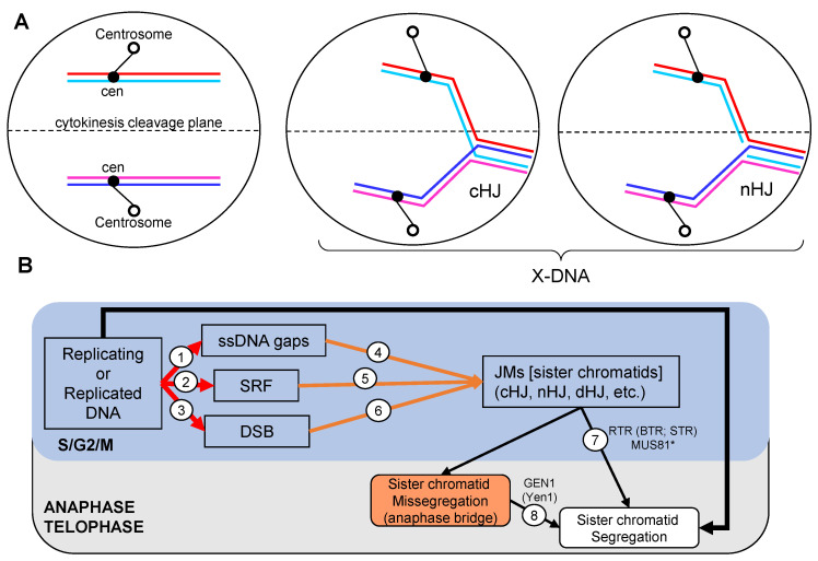
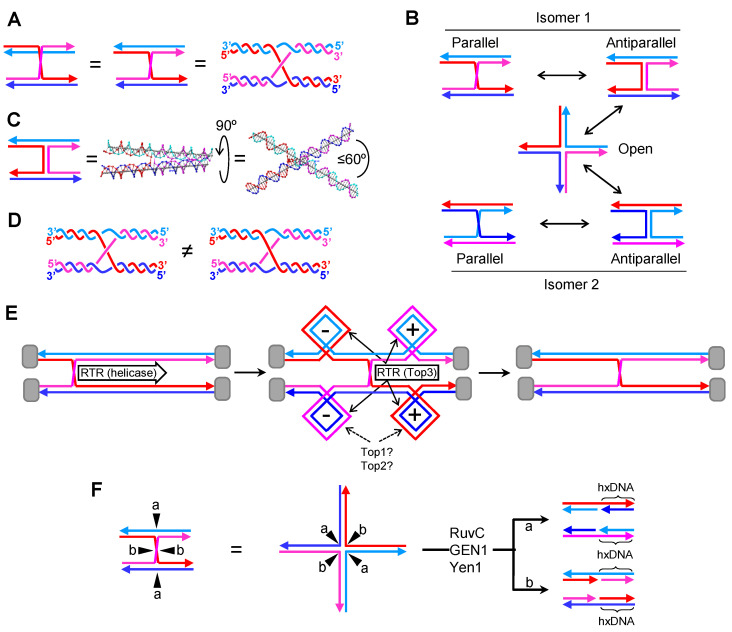
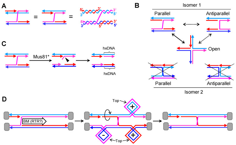
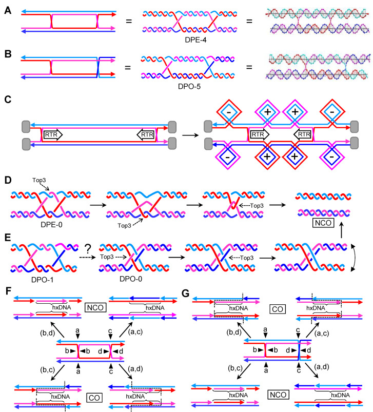
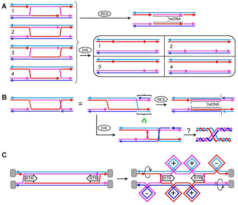
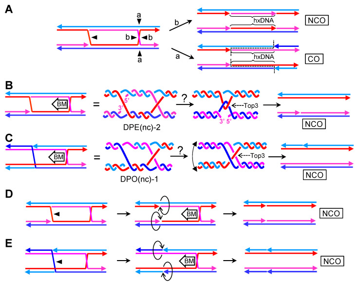
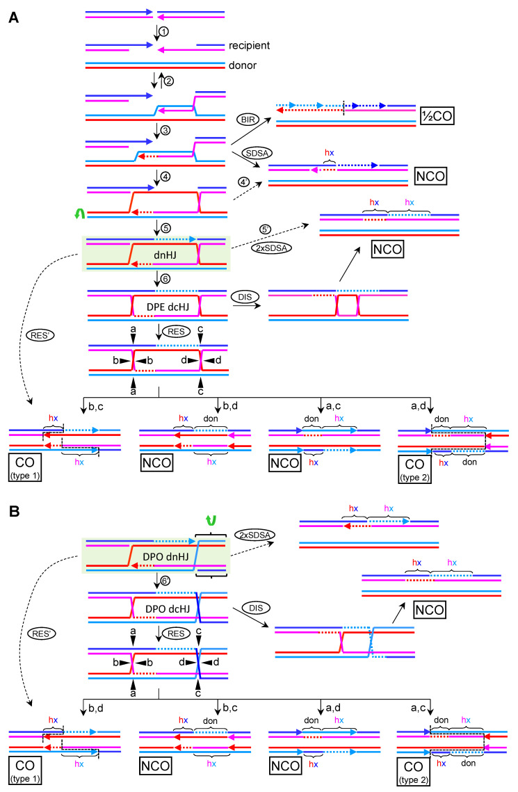
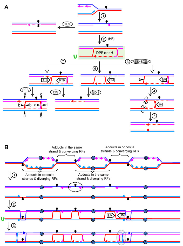
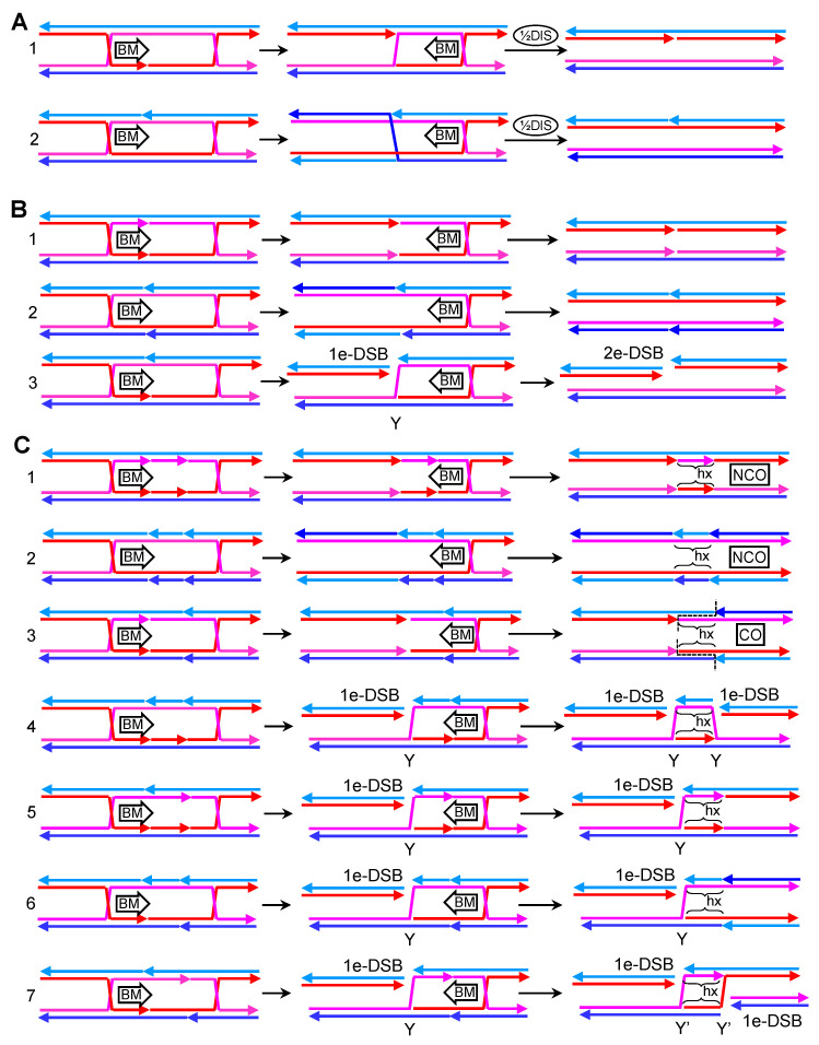
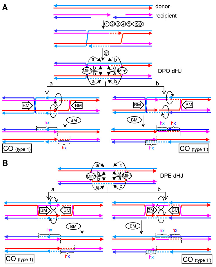
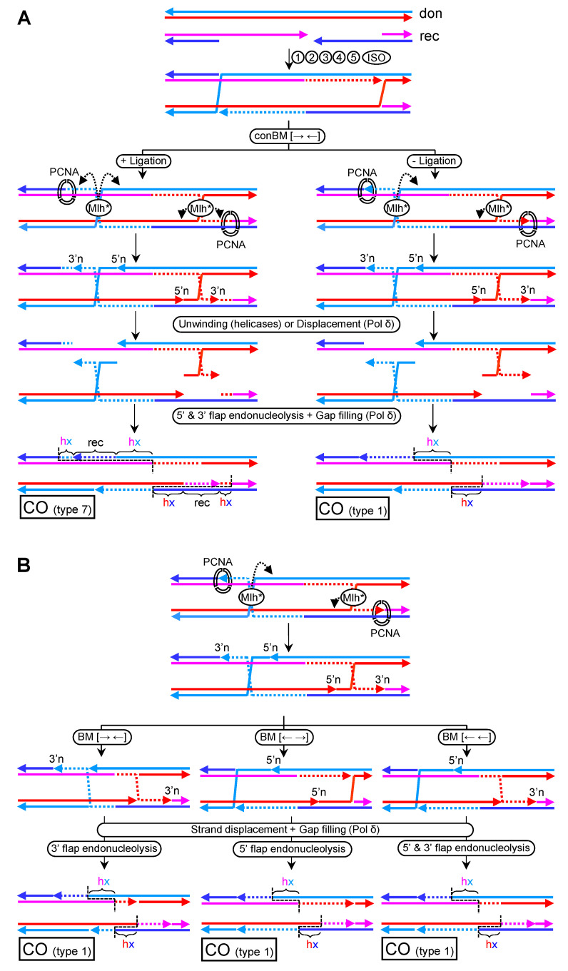
Joint molecules (JMs) are intermediates of homologous recombination (HR). JMs rejoin sister or homolog chromosomes and must be removed timely to allow segregation in anaphase. Current models pinpoint Holliday junctions (HJs) as a central JM. The canonical HJ (cHJ) is a four-way DNA that needs specialized nucleases, a.k.a. resolvases, to resolve into two DNA molecules. Alternatively, a helicase-topoisomerase complex can deal with pairs of cHJs in the dissolution pathway. Aside from cHJs, HJs with a nick at the junction (nicked HJ; nHJ) can be found in vivo and are extremely good substrates for resolvases in vitro. Despite these findings, nHJs have been neglected as intermediates in HR models. Here, I present a conceptual study on the implications of nicks and nHJs in the final steps of HR. I address this from a biophysical, biochemical, topological, and genetic point of view. My conclusion is that they ease the elimination of JMs while giving genetic directionality to the final products. Additionally, I present an alternative view of the dissolution pathway since the nHJ that results from the second end capture predicts a cross-join isomerization. Finally, I propose that this isomerization nicely explains the strict crossover preference observed in synaptonemal-stabilized JMs in meiosis.

Keywords: BLM/Sgs1; GEN1/Yen1; Mlh1-Mlh3/MutLγ; Mus81; ZMM pathway; dissolution pathway; double strand breaks; holliday junction; homologous recombination; replication stress.


Conflict of interest statement

The author declares no conflict of interest. The funders had no role in the design of the study; in the collection, analyses, or interpretation of data; in the writing of the manuscript, or in the decision to publish the results.

Figures

Figure 1
Figure 1
Joint molecules (JM) interfere with chromosome segregation. (A) Examples of aborted chromosome segregation due to the presence of a single JM on one chromosome arm. I depict the two major X-shaped JMs considered in this work: the canonical Holliday junction (cHJ) and the nicked Holliday junction (nHJ). The lack of resolution of the distal part of the chromosome arm results in anaphase bridges that can be broken during cytokinesis. On the left, faithful segregation is accomplished when no JMs are present. (B) The origin and resolution of JMs in the context of the mitotic cell cycle. Replication stress is common even in an unperturbed cell cycle, leading to single-stranded DNA (ssDNA) gaps (1), stalled replication forks (SRFs) (2), and DNA double strand breaks (DSBs) (3). DSBs may also occur pre- and post-replicatively through several mechanisms. At the time of replication (S-phase) or right afterwards (G2/M), cells can deal with ssDNA gaps, SRFs, and DSBs through specialized DNA repair pathways that depend on the homologous recombination (HR) machinery (4,5,6), which create transient links between two DNA molecules—i.e., JMs. JMs are, in turn, eliminated through several mechanisms that are hierarchically regulated. These include dissolution of double cHJ by the RecQ-like Top3 complex (RTR) in S/G2 (7), and resolution by structure-selective endonucleases (SSE), such as Mus81* in G2/M (7) and GEN1/Yen1 in anaphase (8).
Figure 2
Figure 2
Features of the canonical Holliday junction (cHJ). (A) Classical 2D schematics of a cHJ in the parallel conformation. Left: Watson and Crick strands are depicted as straight lines, with the Watson strand on the top in each molecule. Center: the strands of the upper molecule are vertically flipped so the cross-join is easier to visualize. Right: a plectonemic drawing of the strands whereby the double helix of the DNA can be inferred more easily. (B) The cHJ can exist as two isomers, which depend on the pair of strands that forms the cross-join. Each isomer can adopt two extreme conformations, parallel and antiparallel. An open planar conformation serves as an intermediate between isomers and conformers. (C) The antiparallel conformation with tilted arms in the most thermodynamically favored stereoisomer. The antiparallel conformer is depicted as straight lines (left), or modelled with Nanoengineer-1 on the same XY plane (center) and after turning 90° on the Z-plane (right). (D) The spatial position of the strands in the cross-join results in different topoisomers. On the left, the Watson strand of the upper molecule crosses under the Watson strand of the lower molecule. On the right, the crossing plane is inverted. (E) Branch migration in topologically constrained molecule ends (grey boxes) results in positive (+) supercoiling ahead and negative (−) supercoiling behind. The putative activities of the RTR complex in each step are indicated (helicase or topoisomerase). Top3 could deal with supercoiling alone or with the aid of Top1 and/or Top2. (F) The Holliday junction is removed by resolvases by a nicking and counter-nicking mechanism. Two cutting planes are feasible (“a” and “b”). In the open conformation, each plane should have the same chance, so that two distinct products are equally possible. Resolution of a single cHJ gives rise to two new molecules with one flank identical to the parental DNAs and the other flank with cross annealing of Watson and Crick strands coming from different parental DNAs—i.e., heteroduplex DNA (hxDNA). Curly brackets mark out the hxDNA region in each product. Blueish lines, Crick strands; reddish lines, Watson strands. Dark (blue, red) and light (cyan, pink) colors are also included so that the four strands can be differentiated. Parental dsDNA anneals a light and a dark strand, either cyan with red or pink with blue; whereas hxDNA anneals either light–light (cyan–pink) or dark–dark (red–blue) strands. The arrowhead indicates the 3′ end. The equal sign links different visual schematics of the same molecule.
Figure 3
Figure 3
Features of the nicked Holliday junction (nHJ). (A) 2D schematics of the nHJ in the parallel conformation. (B) The nHJ can exist as one isomer, in which the crossover strand corresponds to the homolog to that with the nick. The nHJ tends to adopt the open conformation. (C) The nHJ is the target of a specialized nHJ resolvase, Mus81*, which cuts the crossover strand near the cross-join, leading to two daughter molecules, one with a 5′ flap and one with a gap. The flap and the gap can be further processed in vivo, so the resulting molecules are like one of the two solutions shown in Figure 2F, including the hxDNA flank. (D) Branch migration can isomerize a nHJ into a cHJ. In topologically constrained molecule ends (grey boxes), branch migration results in positive supercoiling (+) ahead and negative supercoiling (−) behind the migration direction. In principle, the nick left behind in one strand may relieve negative supercoiling through swirling (turning arrow). For other details see the legend of Figure 2. BM, Branch migration.
Figure 4
Figure 4
Features of the double canonical Holliday junction (dcHJ). (A) Classical 2D schematics of the dcHJ depicted in DSBR models. The double cross-joins involve the same two strands (Watson strands in this example). This kind of dcHJ spans an even number of half helical turns. The schematics depict the parallel conformation with 4 helical half turns between the cHJs (DPE-4; D stands for dHJ, P for parallel conformation and E for even number of helical half turns). (B) 2D schematics of an dcHJ isomer where each cHJ has a different cross-join partner (Watson strands in the first cHJ and Crick stands in the second cHJ). This isomer changes the number of helical half-turns to an odd number; in this example, there are 5 half-turns (DPO-5; O stands for odd). (C) Converging branch migration of dcHJs results in twice more positive (+) and negative (−) supercoiling than migrating a single cHJ the same distance. (D) Converging Branch migration of a DPE dcHJ to zero half-turns gives rise to hemicatenanes that tie the four strands as in a sailing knot. The complexity of the knot depends on the cross-join topology (Figure 2D). Top3 can unlink the strands as in the example so that the dcHJ is dissolved. (E) Converging Branch migration of a DPO dcHJ to zero would give rise to a topologically distinct sailor knot. To reach a DPO-0, branch migration should take place in steps of half-turns or less, so that the DPO-1 to DPO-0 transition is feasible. (F) The resolution of the DPE dcHJ and its genetic outcomes. Depending on the cutting planes, the resolution can end up in products with or without a terminal genetic exchange—i.e., crossover (CO) or non-crossover (NCO), respectively. Regardless of the product, resolution results in hxDNA between the cHJs (regions with brackets, and also dash lines in COs). (G) The resolution and genetic outcomes of the DPO dcHJ are similar than the DPE dcHJ, albeit with swapped NCO/CO products for the indicated cutting planes. For other details see previous figure legends.
Figure 5
Figure 5
Features of the double nicked Holliday junction (dnHJ). (A) 2D straight line schematics of four DPE dnHJ isomers comprising different combinations of nicked strands. In isomer 1 and 2, one out of the four strands carries two nicks. In isomers 3 and 4, Watson strands carry one nick each. All isomers lead to a single resolution (RES) NCO product with hxDNA between the resolved nHJs. In addition, a single pattern of four nicks is expected. Dissolution (DIS) of all isomers lead to NCOs without hxDNA. The relative position of the two nicks is distinct and may serve as a footprint to infer the original dnHJ. (B) Two-dimensional schematics of a DPO dnHJ isomer. Since the nHJ isomer with the nick in one of the non-crossover strands is highly unfavorable, the more accurate representation is the one shown in the center, with the right edge vertically flipped to represent the single strand in the cross-join (brackets with the green arched arrow). Resolution leads to a single CO product with hxDNA between the resolved nHJs. Dissolution suffers from the problems stated in Figure 4E. (C) Converging Branch migration of a dnHJ would lead to dcHJ with the same positive supercoiling issues raised in Figure 4C. However, negative supercoiling could be relieved as a result of the strand swirling at the nicks left behind. For other details see previous figure legends.
Figure 6
Figure 6
Features of a double Holliday junction formed by a canonical and a nicked Holliday junction (dcnHJ). (A) Resolution of a DPE dcnHJ isomer. The NCO or CO product depends on the cutting plane of the cHJ (“a” or “b”). In both cases a short-tract of hxDNA spans between the resolved HJs. (B) Dissolution of a DPE dcnHJ by branch migration of the cHJ towards the nHJ. A final Top3-mediated ssDNA decatenation reaction appears necessary. An NCO product without hxDNA and with one single nicked strand is expected. (C) Dissolution of a DPO dcnHJ by branch migration of the cHJ towards the nHJ. Top3-mediated ssDNA decatenation could also be necessary. A similar NCO product is expected. (D,E) The dcnHJ can be eliminated through nHJ resolution (black arrowhead) followed by branch migration of the cHJ towards the resulting two opposing nicks. Branch migration might be favored by the absence of positive supercoiling ahead because nicks would enable strands swirling (arched arrows). The NCO product carries two opposing nicks, footprinting the original strands involved in the cross-join. A DPE dcnHJ is shown in (D), and a DPO dcnHJ in (E).
Figure 7
Figure 7
HR pathways for DSB repair. (A) Canonical model with DSBR through DPE dHJs. Steps: (1) Resection of DSB 5′ ends; (2) one 3′ overhang invades a donor homologous sequence to form a reversible D-loop; (3) the D-loop primes de novo DNA synthesis (red dotted line); (4) the second DSB end is captured by the displaced strand; (5) the second capture also primes de novo DNA synthesis (cyan dotted line), and when synthesis from both ends cover the resected tracts, a dnHJ is formed; (6) ligation of the nicks results in a DPE dcHJ; (DIS) dissolution of the dcHJ by RTR results in NCOs with two-sided heteroduplex DNA in the recipient molecule; (RES) resolution by SSEs can result in four different products, two NCOs and two COs, with different local genetic patterns (see main text for further details). Deviations from the DSBR model: (BIR) DNA synthesis from the D-loop continues until the end of the chromosome, lagging-type synthesis makes the complementary strand; (SDSA and 4′) the D-loop is dismantled after some DNA synthesis, the extended strand reanneals with its parental complementary strand and further DNA synthesis fills in the ssDNA gaps; (2xSDSA/5′) the dnHJ is disassembled and strands reanneal with their parental counterparts; (RES’) SSEs cut the dnHJ resulting in just a single resolution product (type 1 CO). (B) The alternative DSBR model with DPO dHJs. This model includes the cross-join isomerization expected for the second nHJ, resulting in a DPO dnHJ; (6′) ligation would yield a DPO dcHJ. From a genetic point of view, the products of SSE resolution (RES and RES’), RTR dissolution (DIS), and dnHJ disassembly (2xSDSA) are identical to those of DPE dHJs. Other subpathway abbreviations: BIR, break-induced replication; SDSA, synthesis dependent strand annealing. Abbreviations for global genetic products: NCO, non-crossovers; CO, crossovers; ½CO, half crossovers. Abbreviations for local genetic rearrangements: hx, heteroduplex DNA tract (delimited by brackets; the color of each letter encodes the strands involved in the heteroduplex); don, donor sequence tract. The green arched arrow indicates a visual flipping of DNA molecules, or part of them (brackets indicate the flipping portion). The black arrowheads and attached lower case letters point to the nicking planes during resolution. For other abbreviations and signs see previous figure legends.
Figure 8
Figure 8
Bypass of replication blockage. (A) Gap repair during RS: (1), a DNA adduct (black bulb) blocks synthesis of the lagging strand during replication, leaving a ssDNA gap behind the RF; (2) the 3′ end melts and invades the sister chromatid, which acts as a donor to prime DNA synthesis and bypass the blockage, while the displaced strand is captured by the ssDNA gap, resulting in a DPE dncHJ; (3–5) the JM is processed by first resolving the nHJ with an SSE, followed by branch migration of the cHJ towards the opposing nicks (RES+SDSA-like); (6) alternatively, the cHJ could migrate towards the nHJ and both are eliminated through a half dissolution pathway (½DIS); (7) the nHJ is ligated into a cHJ, and the resulting DPE dcHJ is processed as in Figure 7. The alternative non-HR translesion synthesis pathway (TLS) is also depicted. (B) DPO dcHJ can emerge through branch migration from two contiguous DPE dHJs. DNA adducts are expected to spread along a replicating DNA molecule. Several combinations regarding the relative position of adducts, direction of RFs and leading/lagging strand blockage are possible (indicated with brackets). (1) Replication termination would leave ssDNA gaps, even for leading strand blockage (dotted circle); (2) gap repair would proceed as in panel A to yield tandem dncHJs, which can be orientated as nHJ-cHJ-nHJ-cHJ (head-to-tail), cHJ-nHJ-nHJ-cHJ (head-to-head) or nHJ-cHJ-cHJ-nHJ (tail-to-tail); (3) Diverging branch migration can put in proximity two cHJ coming from different bypass events, resulting in DPO dcHJs when two adjacent dncHJs where in either head-to-head or tail-to-tail orientation (grey oval for an example coming from a tail-to-tail partner). An “O” within a dark blue circle points to replication origins. For other abbreviations and signs see previous figure legends.
Figure 9
Figure 9
Inferences of metastable nicks during branch migration of a DPE dcHJ. (A) Scenarios with one nick: (1) in a crossover strand; (2) in a non-crossover strand. (B) Scenarios with two opposing nicks: (1) in the crossover strands; (2) in the non-crossover strands; (3) in one crossover and one non-crossover strand. (C) Scenarios with two partners of opposing nicks: (1) all four nicks in the crossover strands; (2) all four nicks in the non-crossover strands; (3) one partner in the crossover strands and the other one in the non-crossover strands; (4) both partners in a crossover/non-crossover arrangement, with only two affected strands; (5) one partner in a crossover/non-crossover arrangement, and the other one in the crossover strands; (6) one partner in a crossover/non-crossover arrangement, and the other one in the non-crossover strands; (7) both partners in a crossover/non-crossover arrangement, with all four strands holding a nick. 1e-DSB, one-ended DSB; 2e-DSB, two-ended DSB; Y and Y’, RF-like structures. Y-Y partner resembles a replication bubble, whereas the Y’-Y’ partner makes a bubble-like JM that connects a CO-to-be chimera. See previous figure legends for other abbreviations and signs.
Figure 10
Figure 10
Models for type 1 CO specificity of ZMM meiotic resolution based on Mlh1-Mlh3 making incisions in trans. (A) Inward nicking by Mlh1-Mlh3 upon a DPO dHJ. Steps (1–5) are like in Figure 7A, and includes the nHJ isomerization (ISO) of Figure 7B. In step 6′, Mlh1-Mlh3 polymerizes inwards from the cHJs/nHJs (cHJs in this example). The main restriction of the model is that incisions in trans takes place with strand specificity relative to the nucleating HJ: “a” for crossover strands and “b” for non-crossover strands; “a” is also compatible with targeting first incisions to the newly synthesized strand sections. Branch migration towards the opposing nicks results in the resolution of the cHJs. (B) First incisions targeted by the newly synthesized strand section also predict type 1 COs in DPE dcHJ.
Figure 11
Figure 11
Models for ZMM meiotic resolution based on Mlh1-Mlh3 making incisions in cis upon a DPO dHJ. (A) Models in which cHJs are resolved from flanking nicks in the crossover strands. Steps 1–5 and ISO are like in Figure 10. Then, convergent branch migration is needed to relocate the newly synthesized tracts within the cross-joins, so that PCNA directs incisions into the crossover strands. Several mechanisms can then process each cHJ from the new nick located 5′ to the cHJ (5′n). To ease visualization, two steps are depicted. Firstly, the unwinding and separation of the portion of nicked crossover strands that stretched across the cHJ, which resolves the cHJ into a 5′-flap and a 3′-flap. This can be achieved by the concerted actions of 3′→5′ and 5′→3′ helicases, or the displacement activity of Pol δ. Secondly, the resulting ssDNA gap is filled in by Pol δ, and the flaps are removed by specialized flap endonucleases. Note that strand displacement and gap filling occur in a single step when Pol δ commences resolution. If a DPO dcHJ is the substrate (left branch; +ligation), a double incision per cHJ is required. Because of the polarity of PCNA, the incision located 3′ to the cHJ (3′n) ought to occur within the newly synthesized track. Thus, the genetic outcome is a reciprocal CO with a hx-recipient-hx tract (type 7). If a DPO dnHJ was the substrate before the converging migration (right branch; -ligation), only the 5′n incision is needed and the mandatory genetic outcome is a type 1 CO, since the 3′n coincides with the end of the HR-driven synthesis by Pol δ. (B) Variations of the right branch of A where branch migration towards one of the nicks precedes the action of Pol δ (or helicases; not depicted). Branch migration can be convergent towards the 5′ns (left branch), divergent towards the 3′ns (center) or codirectional (right branch). In all cases, DPO dnHJ are the intermediate metabolites and type 1 COs are the genetic outcome.

Similar articles

Cited by

References

    1. San-Segundo P.A. Clemente-Blanco Resolvases, Dissolvases, and Helicases in Homologous Recombination: Clearing the Road for Chromosome Segregation. Genes. 2020;11:71. doi: 10.3390/genes11010071. - DOI - PMC - PubMed
    1. Falquet B., Rass U. Structure-specific endonucleases and the resolution of chromosome underreplication. Genes. 2019;10:232. doi: 10.3390/genes10030232. - DOI - PMC - PubMed
    1. Wright W.D., Shah S.S., Heyer W.D. Homologous recombination and the repair of DNA double-strand breaks. J. Biol. Chem. 2018;293:10524–10535. doi: 10.1074/jbc.TM118.000372. - DOI - PMC - PubMed
    1. Dehé P.-M., Gaillard P.-H.L. Control of structure-specific endonucleases to maintain genome stability. Nat. Rev. Mol. Cell Biol. 2017;18:315–330. doi: 10.1038/nrm.2016.177. - DOI - PubMed
    1. Watt P.M., Louis E.J., Borts R.H., Hickson I.D. Sgs1: A eukaryotic homolog of E. coli RecQ that interacts with topoisomerase II in vivo and is required for faithful chromosome segregation. Cell. 1995;81:253–260. - PubMed

Publication types

LinkOut - more resources