. 2020 Dec 12;12(12):1431.
doi: 10.3390/v12121431.

Bovine Adenovirus-3 Tropism for Bovine Leukocyte Sub-Populations

Affiliations

Bovine Adenovirus-3 Tropism for Bovine Leukocyte Sub-Populations

Sugandhika Khosa et al. Viruses. .

Abstract

A number of characteristics including lack of virulence and the ability to grow to high titers, have made bovine adenovirus-3 (BAdV-3) a vector of choice for further development as a vaccine-delivery vehicle for cattle. Despite the importance of blood leukocytes, including dendritic cells (DC), in the induction of protective immune responses, little is known about the interaction between BAdV-3 and bovine blood leukocytes. Here, we demonstrate that compared to other leukocytes, bovine blood monocytes and neutrophils are significantly transduced by BAdV404a (BAdV-3, expressing enhanced yellow green fluorescent protein [EYFP]) at a MOI of 1-5 without a significant difference in the mean fluorescence of EYFP expression. Moreover, though expression of some BAdV-3-specific proteins was observed, no progeny virions were detected in the transduced monocytes or neutrophils. Interestingly, addition of the "RGD" motif at the C-terminus of BAdV-3 minor capsid protein pIX (BAV888) enhanced the ability of the virus to enter the monocytes without altering the tropism of BAdV-3. The increased uptake of BAV888 by monocytes was associated with a significant increase in viral genome copies and the abundance of EYFP and BAdV-3 19K transcripts compared to BAdV404a-transduced monocytes. Our results suggest that BAdV-3 efficiently transduces monocytes and neutrophils in the absence of viral replication. Moreover, RGD-modified capsid significantly increases vector uptake without affecting the initial interaction with monocytes.

Keywords: EYFP; RGD motif; bovine adenovirus-3; chimeric pIX; leukocytes; tropism.


Conflict of interest statement

The authors declare no conflict of interest. Moreover, the funders had no role in the design of the study; in the collection, analyses, or interpretation of data; in the writing of the manuscript, or in the decision to publish the results.

Figures

Figure 1
Figure 1
Transduction of bovine cells by BAV304a. (A) Schematic diagram of BAV304a genome. Human cytomegalovirus immediate early gene promoter (CMV) and enhanced yellow fluorescent protein gene (EYFP) were inserted in Early (E)-3 region. The direction of transcription is shown by an arrow. Dashed line represents deleted region. The name of the plasmid is on the left and name of the virus is indicated on the right. Purified bovine peripheral blood mononuclear cells (PBMCs) (B) or purified bovine PMNs (C) were infected with increasing MOI of CsCl purified BAV304a. At 18 h post infection, the cells were collected and analyzed to quantify the percentage of cells expressing EYFP (subpanel 1) and the mean florescent intensity (MFI) of EYFP expression in transduced cells (subpanel 2).
Figure 2
Figure 2
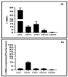
Transduction of leukocyte subpopulations by BAV304a. (A) Bovine PBMCs were isolated and labeled to quantify the percentage of cells within major leukocyte lineages: T cells (CD3+); monocytes (CD14+), B cells (CD21+); innate lymphoid cells (CD335+), and dendritic cells (CD209+). Data presented are the mean + 1 SD of values for PBMCs isolated from five animals. (B) Bovine PBMCs were transduced with BAV304a (MOI 2) for 18 h before labelling with leukocyte lineage-specific monoclonal antibodies and analyzed for co-expression of EYFP. Data presented are the mean + 1 SD of the percentage of cells within each leukocyte lineage expressing EYFP. Transduction experiment was repeated with PBMCs isolated from five animals.
Figure 3
Figure 3
Replication of BAV304a in monocytes and PMNs. Purified CD14+ bovine monocytes (A) or bovine PMN cells (C) were infected with BAV304a at an MOI of 2. At 48 h post-infection, the cells were examined by light microscopy (Trans) to confirm viability and with fluorescent microscopy (EYFP expression) to confirm transduction. The infected cells were collected, freeze-thawed and the virus in infected monocyte lysates (B) or infected PMNs lysates (D) was titrated using MDBK cells. Viral infection of MDBK cells was analyzed for cytopathic effect (CPE) with light microscopy (Trans) and expression of EYFP by fluorescent microscopy (EYFP expression). Results presented are representative of two independent experiments with three replicates cultures in each experiment. Five-fold serial dilutions of monocyte lysates and two-fold serial dilutions of PMN lysates were used for virus titration on MDBK cells. Magnification in all images is 100×.
Figure 4
Figure 4
Viral protein expression in BAV304a-infected cells. Monocytes (A) and PMNs (B). The infected cell lysates were separated with 12% SDS-PAGE, transferred to nitrocellulose membranes and probed in Western blot using BAdV-3 anti-19K serum (subpanel 1), BAdV-3 anti DBP serum (subpanel 2), BAdV-3 anti-52Kserum (subpanel 3), BAdV-3 anti-hexon serum (subpanel 4), and BAdV-3 anti-100K serum (subpanel 5). BAV304a-infected MDBK cells (panel A, lanes 1; panel B lanes 2); BAV304a-infected monocytes (panel A, lanes 3) or PMNs (panel B, lanes 2); mock-infected monocytes (panel A, lanes 2) or PMNs (panel B, lanes 1); molecular weight markers (M) in kDa are shown on the left.
Figure 5
Figure 5
Construction and characterization of BAV888. (A) Schematic diagram of BAV304a and BAV888 genomes; cathepsin cleavage site (CCS); 10 repeats of glycine-serine (spacer), RGD amino acids (RGD). The shaded box represents the BAdV-3 genome sequence, while the dashed line in early (E)-3 region represents deletion in this region. Human cytomegalovirus immediate early gene promoter (CMV); enhanced yellow fluorescent protein (EYFP) gene. The direction of transcription is indicated with arrows. The name of each plasmid is indicated to the left and virus name is indicated to the right. (B) EYFP fluorescence. The cells were infected with BAV888 and analyzed by Leica TC5 SP5 immunofluorescence microscope (Magnification 400×). (C) Expression of pIX in BAV304a and BAV888 infected cells. The proteins from the lysates of mock-infected cells (lane 1), BAV304a infected cells (lane 2) or BAV888 infected cells (lane 3) were separated with 15% SDS-PAGE, transferred to a nitrocellulose membrane and probed in Western blot using anti-pIX antiserum. The pIX protein is indicated by arrows. Molecular weight markers (M).
Figure 6
Figure 6
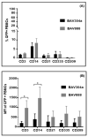
Comparison of bovine leukocyte transduction by BAV304a or BAV888. Bovine PBMCs were transduced for 16 h with either BAV304a or BAV888 (MOI = 1). Cells were labelled with lineage-specific monoclonal antibodies and analyzed for co-expression of GFP: T cells (CD3+), monocytes (CD14+), B cells (CD121+), innate lymphoid cells (CD335+), and DCs (CD209+). The percent GFP+ cells (panel A) and EYFP mean fluorescence intensity (MFI) (panel B) were analyzed for each PBMC lineage. Data presented are the mean + 1SD of values from PBMCs isolated from five animals. * Significant (p < 0.05) differences when comparing BAV304a or BAV888 transduction within a leukocyte lineage.
Figure 7
Figure 7
BAdV-3 genome copy number and viral gene expression in bovine monocytes. Monocytes (CD14+ cells) were isolated from PBMCs using MACS and transduced with either BAV304a or BAV888 using an MOI of 1. After 16 h, cells were collected and DNA and RNA were extracted. Total viral genome copy number recovered from each culture was quantified by qPCR (A). Expression of the EYFP gene and both early and late viral genes was quantified with qRT-PCR. Ct values for genes were normalized with β-actin (ΔCt) and presented as 1/ΔCt and compared for BAV304a- and BAV888-infected monocytes (B). Relative expression of EYFP and individual viral genes was compared for BAV304a (C) and BAV888 (D). Data presented are the mean + 1SD of values from three independent r experiments. * (p < 0.05); ** (p < 0.01); *** (p < 0.001); **** (p < 0.0001).

Similar articles

References

    1. Tatsis N., Ertl H.C. Adenoviruses as vaccine vectors. Mol. Ther. 2004;10:616–629. doi: 10.1016/j.ymthe.2004.07.013. - DOI - PMC - PubMed
    1. Crystal R.G. Transfer of genes to humans: Early lessons and obstacles to success. Science. 1995;270:404–410. doi: 10.1126/science.270.5235.404. - DOI - PubMed
    1. Ayalew L.E., Pankaj Kumar P., Gaba A., Makadiya N., Tikoo S.K. Bovine adenovirus-3 as a vaccine delivery vehicle. Vaccine. 2015;33:493–499. doi: 10.1016/j.vaccine.2014.11.055. - DOI - PMC - PubMed
    1. Lehmkuhl H.D., Smith M.H., Dierks R.E. A bovine adenovirus type 3: Isolation, characterization and experimental infection in calves. Arch. Virol. 1975;48:39–46. doi: 10.1007/BF01320564. - DOI - PubMed
    1. Baxi M.K., Deregt D., Robertson J., Babiuk L.A., Schlapp T., Tikoo S.K. Recombinant bovine adenovirus type 3 expressing bovine viral diarrhea virus glycoprotein E2 induces an immune response in cotton rats. Virology. 2000;278:234–243. doi: 10.1006/viro.2000.0661. - DOI - PubMed

Publication types

Supplementary concepts

LinkOut - more resources