. 2021 May;28(5):1720-1732.
doi: 10.1038/s41418-020-00698-4. Epub 2020 Dec 16.

Gender biased neuroprotective effect of Transferrin Receptor 2 deletion in multiple models of Parkinson's disease

Affiliations

Gender biased neuroprotective effect of Transferrin Receptor 2 deletion in multiple models of Parkinson's disease

Chiara Milanese et al. Cell Death Differ. 2021 May.

Abstract

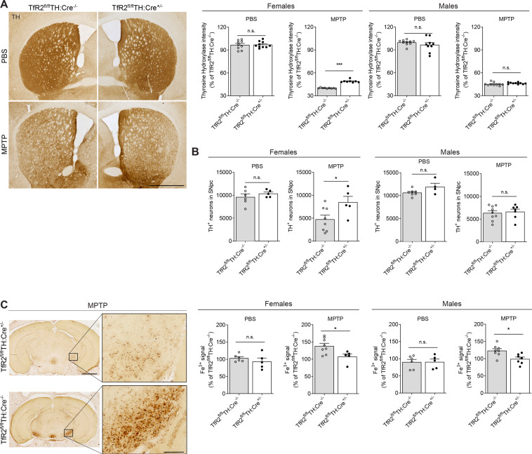
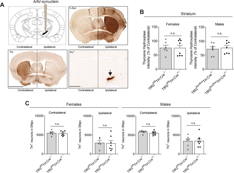
Alterations in the metabolism of iron and its accumulation in the substantia nigra pars compacta accompany the pathogenesis of Parkinson's disease (PD). Changes in iron homeostasis also occur during aging, which constitutes a PD major risk factor. As such, mitigation of iron overload via chelation strategies has been considered a plausible disease modifying approach. Iron chelation, however, is imperfect because of general undesired side effects and lack of specificity; more effective approaches would rely on targeting distinctive pathways responsible for iron overload in brain regions relevant to PD and, in particular, the substantia nigra. We have previously demonstrated that the Transferrin/Transferrin Receptor 2 (TfR2) iron import mechanism functions in nigral dopaminergic neurons, is perturbed in PD models and patients, and therefore constitutes a potential therapeutic target to halt iron accumulation. To validate this hypothesis, we generated mice with targeted deletion of TfR2 in dopaminergic neurons. In these animals, we modeled PD with multiple approaches, based either on neurotoxin exposure or alpha-synuclein proteotoxic mechanisms. We found that TfR2 deletion can provide neuroprotection against dopaminergic degeneration, and against PD- and aging-related iron overload. The effects, however, were significantly more pronounced in females rather than in males. Our data indicate that the TfR2 iron import pathway represents an amenable strategy to hamper PD progression. Data also suggest, however, that therapeutic strategies targeting TfR2 should consider a potential sexual dimorphism in neuroprotective response.


Conflict of interest statement

The authors declare that they have no conflict of interest.

Figures

Fig. 1
Fig. 1. Targeted TfR2 deletion in TH expressing cells and PD modeling in mice.
A PCR to confirm TfR2 deletion performed on genomic DNA extracted from 30-μm-thick PFA-fixed sections of the olfactory bulbs, which abound in TH-neurons. DNA from two consecutive slices was used in each PCR reaction. Genotyping was performed using two different sets of primers, one of which (TfR2) amplifies the WT or floxed allele, while the other (TfR2 deleted) amplifies the deleted allele. Selective recombination of TfR2 selectively occurs only in Cre-expressing floxed mice (Tfr2fl/flTH:Cre+/−, last lane) and not in mice lacking Cre expression (lanes 2 and 4). B Validation of the PD-related treatments used in the study, which successfully elicited dopaminergic degeneration, as indicated by unbiased stereological counts. Graphs represent mean + SEM, two-tailed Student’s t test; *p < 0.05, **p < 0.005, ***p < 0.001.
Fig. 2
Fig. 2. TfR2 deletion is protective in 12-week-old MPTP-treated mice.
A TH immunoreactivity in striata of MPTP or vehicle-treated animals. TfR2 deletion mitigates striatal denervation in brains of female mice. B Unbiased stereological counts of TH immunopositive neurons in the substantia nigra confirm that TfR2 targeted deletion is protective in 12-week-old females, but not in males. C Perls’ staining for Fe3+ reveals reduced iron accumulation in the midbrain after MPTP treatment in animals with TfR2 targeted deletion (12-week-old mice). Data in experimental groups are expressed as percentage of the respective TfR2fl/flTH:Cre−/− vehicle-treated control group. Two-tailed Student’s t test; *p < 0.05, **p < 0.005, ***p < 0.001. Calibration bars: 1 mm in (A), 2 mm (left) and 100 μm (right) in (C).
Fig. 3
Fig. 3. TfR2 deletion is protective in 52-week-old MPTP-treated mice.
A Striatal TH immunoreactivity in 52-week-old mice. TfR2 deletion provides moderate, yet significant protection from MPTP treatment. B Unbiased stereological counts of TH immunopositive neurons in the SNpc confirm that TfR2 deletion is protective in females, but not in males. C. Perls’ staining for Fe3+ reveals significant decrease in iron accumulation in the substantia nigra in after MPTP treatment in animals with TfR2 targeted deletion. In 52-week-old mice, TfR2 deletion also reduces accumulation of iron deposits in untreated females. D Double histological staining to reveal Fe3+ in TH immunopositive neurons in the SNpc, in 12- and 52-week-old mice. Iron deposits partially co-localize with dopaminergic neurons (arrows). Iron levels are remarkably more evident in 52-week than in 12-week-old-treated animals. All data are expressed as percentage of the TfR2fl/flTH:Cre−/− vehicle-treated control group. Two-tailed Student’s t test; *p < 0.05, **p < 0.005, ***p < 0.001. Calibration bars: 1 mm in (A), 2 mm in (C) and 50 μm in (D).
Fig. 4
Fig. 4. Effects of TfR2-targeted deletion in an AAV-h-alpha-synuclein overexpressing model of PD.
A Schematic of intracranial injection of AAV-h-synuclein in the right SNpc hemisphere (ipsilateral). Transduction with rAAV coding for human alpha-synuclein (h-syn) results in its striatal increase in the ipsilateral hemisphere, as showed in the top left panel that is paralleled by decreased TH levels (bottom left panel) and remarkable Fe3+ deposits accumulation at the site of injection in the SNpc, indicative of ongoing pathology. B, C TfR2 deletion does not provide any protection against AAV-h-synuclein overexpression, as indicated by striatal dopaminergic innervation expressed as percentage of the contralateral side (B) and unbiased stereological counts of dopaminergic neurons in the SNpc. Graphs represent scatter plots with bars. Two-tailed t test. Calibration bars: 1 mm (top) and 2 mm (bottom) in (A).
Fig. 5
Fig. 5. TfR2-targeted deletion is partially protective against alpha-synuclein pre-formed fibrils (PFF) intracranial seeding.
A Schematic of the stereotactic injection procedure during which PFF have been injected in the right striatum. B TfR2 deletion provides protection against striatal denervation induced by PFF, as indicated by TH immunohistochemistry. C Unbiased stereological counts of dopaminergic neurons in the SNpc indicate that TfR2-targeted deletion is protective against PFF-induced degeneration in females, but not in males. D Perls’ staining for Fe3+ did not reveal any effect of TfR2 deletion on iron accumulation in the SNpc induced by PFF (left panel). Immunodetection of TH-positive neurons combined with Perls’ staining reveals iron deposits also in SNpc dopamine neurons (right panel). E Increased levels of ser129 p-α-synuclein in the substantia nigra (arrows) confirms α-synuclein stress in PFF-injected animals that is not ameliorated by TfR2 deletion (green signal). Graphs represent scatter plots with bars. Two-tailed t test; *p < 0.05, **p < 0.005, ***p < 0.001. Calibration bars: 1 mm in (B), 2 mm (left panel) and 50 μm (right panel) in (D), and 50 μm in (E).

References

    1. Greenamyre JT, Hastings TG. Biomedicine. Parkinson’s-divergent causes, convergent mechanisms. Science. 2004;304:1120–2. - PubMed
    1. Chinta SJ, Lieu CA, Demaria M, Laberge RM, Campisi J, Andersen JK. Environmental stress, ageing and glial cell senescence: a novel mechanistic link to Parkinson’s disease? J Intern Med. 2013;273:429–36. - PMC - PubMed
    1. Collier TJ, Kanaan NM, Kordower JH. Ageing as a primary risk factor for Parkinson’s disease: evidence from studies of non-human primates. Nat Rev Neurosci. 2011;12:359–66. - PMC - PubMed
    1. Surmeier DJ. Determinants of dopaminergic neuron loss in Parkinson’s disease. FEBS J. 2018;285:3657–68. - PMC - PubMed
    1. Gotz ME, Double K, Gerlach M, Youdim MB, Riederer P. The relevance of iron in the pathogenesis of Parkinson’s disease. Ann N Y Acad Sci. 2004;1012:193–208. - PubMed

Publication types