Comparative Study
. 2020 Nov 25:11:575607.
doi: 10.3389/fimmu.2020.575607. eCollection 2020.

Platelets Selectively Regulate the Release of BDNF, But Not That of Its Precursor Protein, proBDNF

Affiliations
Comparative Study

Platelets Selectively Regulate the Release of BDNF, But Not That of Its Precursor Protein, proBDNF

Jessica Le Blanc et al. Front Immunol. .

Abstract

Background: Brain-derived neurotrophic factor (BDNF) plays a role in synaptic plasticity and neuroprotection. BDNF has well-established pro-survival effects, whereas its precursor protein, proBDNF, induces apoptosis. Thus, it has been suggested that the proBDNF/BDNF ratio could be an indicator of neuronal health. Access to neurons is, understandably, limited. Because of their similarities, platelets have been put forward as a non-invasive biomarker of neuronal health; indeed, they store large quantities of BDNF and can release it into circulation upon activation, similarly to neurons. However, whether platelets also express the precursor proBDNF protein remains unknown. We therefore sought to characterize proBDNF levels in human platelets and plasma.

Methods: The presence of proBDNF was assessed by immunoblotting, cell fractionation, flow cytometry, and confocal microscopy in washed platelets from 10 healthy volunteers. Platelets from 20 independent healthy volunteers were activated with several classical agonists and the release of BDNF and proBDNF into plasma was quantified by ELISA.

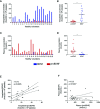
Results: Platelets expressed detectable levels of proBDNF (21 ± 13 fmol/250 x 106 platelets). ProBDNF expression was mainly localized in the intracellular compartment. The proBDNF to BDNF molar ratio was ~1:5 in platelets and 10:1 in plasma. In stark contrast to the release of BDNF during platelet activation, intraplatelet and plasma concentrations of proBDNF remained stable following stimulation with classical platelet agonists, consistent with non-granular expression.

Conclusions: Platelets express both the mature and the precursor form of BDNF. Whether the intraplatelet proBDNF to BDNF ratio could be used as a non-invasive biomarker of cognitive health warrants further investigation.

Keywords: brain-derived neurotrophic factor; plasma; platelets; pro-BDNF; secretion.


Conflict of interest statement

ML has received speaker fees from Bayer; has participated in industry-funded trials from Idorsia; has served on advisory boards for Servier; and has received in-kind and financial support for investigator-initiated grants from Leo Pharma, Roche Diagnostics, Aggredyne, and Fujimori Kogyo. The remaining authors declare that the research was conducted in the absence of any commercial or financial relationships that could be construed as a potential conflict of interest.

Figures

Figure 1
Figure 1
Human platelets contain proBDNF. (A) ProBDNF immunoblotting of human washed platelet lysates (15 µg) from six different healthy volunteers. Recombinant proBDNF (3 ng) and human cortex lysate (3 µg) were used as positive controls. Molecular weight is indicated on the left (kDa) and primary antibody on the right. Experiments representative of n=9 for R-176 and n=10 for mab31751 antibody. IB, immunoblotting. (B) Immunoblotting of proBDNF and BDNF in different fractions of washed human platelets. P-Selectin was used as control protein in the membrane fraction, p65 NF-ĸB was used as control protein in the cytosol, and α-tubulin was used as control protein in the cytoskeleton. The equivalent of the protein content of 3 x 10⁷ platelets was loaded for each fraction on the gel. Representative experiment of n=4 different volunteers. (C) ProBDNF treatment with PNGase F in washed human platelet lysates. U87-MG glioblastoma cells were used as a control. rProBDNF, recombinant proBDNF (3 ng); cortex, human cortex lysate (3 µg); platelets, whole human platelet lysate (representative experiment of n=4 different volunteers, 7.5 x 10⁸ platelets per well); −PNGF, platelets treated with GlycoBuffer, and incubated at 37°C for 60 min without PNGase F; +PNGF, platelets treated with GlycoBuffer and incubated at 37°C for 60 min with PNGase F; PNGF alone, PNGase F incubated at 37°C for 60 min without platelet lysate. CD42b and sortilin were used as controls of protein deglycosylation in platelets and in U87-MG cells, respectively. n=3 different volunteers for PNGase treatments in human platelets and n=4 independent experiments for U87-MG cells. (D) Representative flow cytometry experiment showing surface and intracellular proBDNF in human washed platelets and in U87-MG and U251-MG glioblastoma cell lines. Mouse IgG2b was used as isotype control. Percentage of expression are indicated on the figure. n=10 different healthy volunteers for human platelets; n=3 independent experiments for each glioblastoma cell line. (E) Confocal microscopy imaging of proBDNF in human permeabilized washed platelets (top) and in permeabilized U-251 MG cells (bottom). Mouse IgG2b was used as isotype control. ProBDNF was labelled using Alexa488 fluorochrome (in green). Nuclei were stained with DAPI (in blue). Scale bar = 2 µm and 200 µm for washed platelets and U-251 MG cells images, respectively. (F) Immunoblotting of α2-macroglobulin at increasing quantities of loaded proteins (1–20 µg) obtained from a washed platelet lysate or platelet-poor plasma (PPP) from the same individual (#23). α-tubulin was used as loading control. Molecular weight is indicated on the left (kDa) and primary antibody on the right. PPP, platelet poor plasma; PLTs, platelets; IB, immunoblotting.
Figure 2
Figure 2
Molar concentrations of proBDNF are lower in platelets and higher in plasma than those of BDNF. ELISA quantification of proBDNF and BDNF levels in the intraplatelet (A, B) and plasma (C, D) compartments. Concentrations are normalized for 250 x 106 platelets. Horizontal bar represents median, *p<0.05. (E, F) Correlation between BDNF and proBDNF molar concentrations in human platelets (E) and in plasma (F). Dotted lines represent 95% confidence intervals, n=20 participants (#1 to #20).
Figure 3
Figure 3
Unlike BDNF, intraplatelet proBDNF is not released during platelet activation. Intraplatelet (A, D) and plasma (B, E) concentrations of BDNF and proBDNF following platelet activation by different agonists. Intraplatelet concentrations are normalized for 250 x 106 platelets. Proportion of BDNF (C) and proBDNF (F) in plasma vs. in platelets are expressed in percentage. Error bar represents IQR, *p < 0.05 vs. ctrl, n=20 participants (#1 to #20). Ctrl, control; ADP, adenosine diphosphate; TRAP, thrombin receptor-activating peptide; AA, arachidonic acid.

References

    1. Gibon J, Barker PA. Neurotrophins and Proneurotrophins: Focus on Synaptic Activity and Plasticity in the Brain. Neuroscientist (2017) 23(6):587–604. 10.1177/1073858417697037 - DOI - PubMed
    1. Kowianski P, Lietzau G, Czuba E, Waskow M, Steliga A, Morys J. BDNF: A Key Factor with Multipotent Impact on Brain Signaling and Synaptic Plasticity. Cell Mol Neurobiol (2018) 38(3):579–93. 10.1007/s10571-017-0510-4 - DOI - PMC - PubMed
    1. Foltran RB, Diaz SL. BDNF isoforms: a round trip ticket between neurogenesis and serotonin? J Neurochem (2016) 138(2):204–21. 10.1111/jnc.13658 - DOI - PubMed
    1. Borodinova AA, Salozhin SV. Differences in the Biological Functions of BDNF and proBDNF in the Central Nervous System. Neurosci Behav Physiol (2017) 47(3):251–65. 10.1007/s11055-017-0391-5 - DOI
    1. Teng HK, Teng KK, Lee R, Wright S, Tevar S, Almeida RD, et al. ProBDNF induces neuronal apoptosis via activation of a receptor complex of p75NTR and sortilin. J Neurosci (2005) 25(22):5455–63. 10.1523/JNEUROSCI.5123-04.2005 - DOI - PMC - PubMed

Publication types

Grants and funding

LinkOut - more resources