Review
. 2021 Apr;81(3):253-272.
doi: 10.1002/dneu.22774. Epub 2020 Aug 29.

Tubulin post-translational modifications control neuronal development and functions

Affiliations
Review

Tubulin post-translational modifications control neuronal development and functions

Marie-Jo Moutin et al. Dev Neurobiol. 2021 Apr.

Abstract

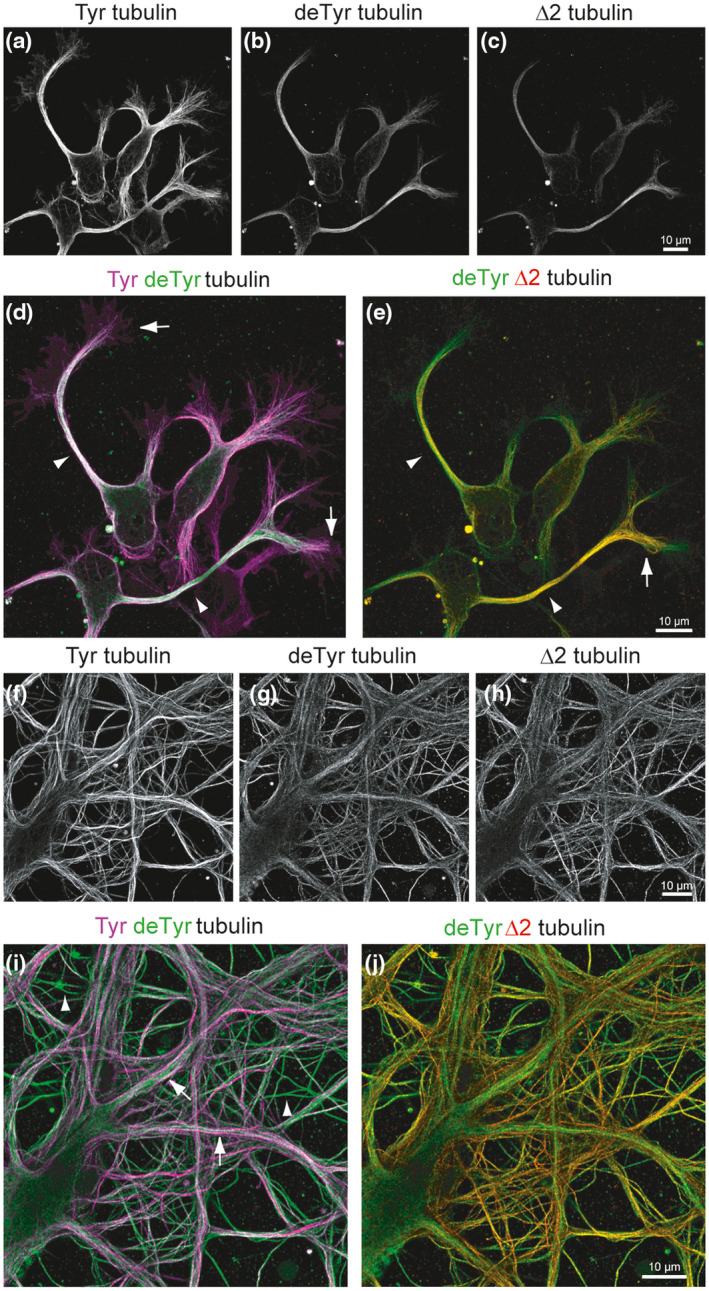
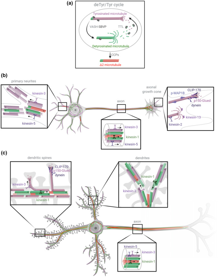
Microtubules (MTs) are an essential component of the neuronal cytoskeleton; they are involved in various aspects of neuron development, maintenance, and functions including polarization, synaptic plasticity, and transport. Neuronal MTs are highly heterogeneous due to the presence of multiple tubulin isotypes and extensive post-translational modifications (PTMs). These PTMs-most notably detyrosination, acetylation, and polyglutamylation-have emerged as important regulators of the neuronal microtubule cytoskeleton. With this review, we summarize what is currently known about the impact of tubulin PTMs on microtubule dynamics, neuronal differentiation, plasticity, and transport as well as on brain function in normal and pathological conditions, in particular during neuro-degeneration. The main therapeutic approaches to neuro-diseases based on the modulation of tubulin PTMs are also summarized. Overall, the review indicates how tubulin PTMs can generate a large number of functionally specialized microtubule sub-networks, each of which is crucial to specific neuronal features.

Keywords: neuro-diseases; neuron; post-translational modifications; tubulin; tyrosination.


Conflict of interest statement

The authors declare no conflict of interest.

Figures

FIGURE 1
FIGURE 1
Distribution of tyrosinated (Tyr), detyrosinated (deTyr), and ∆2 tubulin in developing and mature neurons. Airyscan confocal images of hippocampal neurons after 2 (a‐e) or 18 days in culture (f‐j). Cells were labeled with anti‐Tyr, anti‐deTyr, and anti‐∆2 tubulin antibodies (Aillaud et al., 2016). Static immunostaining images of Tyr, deTyr, and ∆2 tubulin labeling were obtained with a ×63 oil‐immersion objective (1.4 NA) using an inverted confocal microscope (LSM 710, Zeiss) linked to an Airyscan detector to improve signal‐to‐noise ratio and spatial resolution. Scale bar = 10 µm. At early developmental stages, the majority of neuronal microtubules bear Tyr tubulin (a). DeTyr microtubules are enriched in axons (arrowheads), but are also detected in neurites (b). ∆2 tubulin is mainly enriched in axons (c). Color‐combined image of Tyr (magenta) and deTyr tubulin (green) reveals that stable deTyr microtubules are enriched in axons (arrowheads), while most dynamic Tyr microtubules are found in the growth cone (arrows) (d). The color‐combined image of deTyr (green) and ∆2 tubulin (red) reveals a different distribution of these modified tubulins inside the axon: while deTyr and ∆2 tubulin colocalize in axons (arrowheads) and at the proximal part of the axonal growth cone (arrow), deTyr tubulin extends further into neurites and growth cones (e). In mature neurons, dynamic Tyr microtubules are enriched in the outer part of primary large dendrites and in some thin structures that could be axons emanating from neighboring neurons (f). DeTyr microtubules are seen bundled in the inner part of large dendrites, and are also highly enriched in most thin structures (g). ∆2 tubulin is mainly present in the thin structures (h). Color‐combined image of Tyr (magenta) and deTyr tubulin (green) reveals an asymmetric pattern in large dendrites (Tyr outside and deTyr inside, arrowhead), and the enrichment of deTyr microtubules in axon‐like structures (arrow) (i). Color‐combined image of deTyr (green) and ∆2 tubulin (red) reveals a different distribution of the modified tubulins with only partial colocalization in thin axon like structures (j)
FIGURE 2
FIGURE 2
Schematic representation of the three major pools of α‐tubulin resulting from the detyrosination/tyrosination cycle in neurons, with their known effectors. (a) The tubulin cycle and Δ2 tubulin production. The VASH‐SVBP complex (VASH1 or VASH2 associated with small vasohibin‐binding protein, SVBP) removes the C‐terminal tyrosine residue of α‐tubulin incorporated in microtubules to generate detyrosinated (deTyr) microtubules (green). After detyrosinated microtubules depolymerize, tubulin tyrosin ligase (TTL) can once again add a tyrosine residue to α/β tubulin dimers. When these soluble dimers polymerize, they form new tyrosinated (Tyr) microtubules (magenta). In long‐lived detyrosinated microtubules, the penultimate glutamate residue of α‐tubulin can be removed by cytosolic carboxypeptidases (CCPs), generating microtubules composed of Δ2 tubulin (red). (b) Distribution of α‐tubulin pools (Tyr/deTyr/Δ2) in developing neurons. During the development of neurons, primary neurites contain mixed‐polarity microtubules mainly composed of Tyr tubulin (magenta). Stable deTyr microtubules (green) are found in the inner region of neurites. Some neurites also contain hyperstable ∆2 microtubules (red). The axon contains uniform polarity microtubules that are highly enriched in deTyr and ∆2 tubulin, mainly in their inner region. The proximal axonal growth cone is composed of stable deTyr and ∆2 microtubules whereas the dynamic axonal growth cone contains Tyr microtubules in its distal region. (c) Distribution of α‐tubulin pools (Tyr/deTyr/Δ2) in mature neurons. In fully developed neurons, dendrites contain mixed‐polarity microtubules composed of Tyr tubulin in their outer region, and deTyr and ∆2 tubulin in their inner region. The Tyr microtubules from dendrites can transiently enter into dendritic spines. Axonal microtubules have uniform polarity and are mainly composed of stable deTyr and ∆2 tubulin. Molecular effectors of the cycle, such as molecular motors, CAP‐Gly plus‐end proteins (CLIP‐170 and p150 Glued), and MAP1B (phosphorylated) are represented. Figure created with Biorender.com

References

    1. Adamopoulos, A. , Landskron, L. , Heidebrecht, T. , Tsakou, F. , Bleijerveld, O. B. , Altelaar, M. , … Perrakis, A. (2019). Crystal structure of the tubulin tyrosine carboxypeptidase complex VASH1‐SVBP. Nature Structural & Molecular Biology, 26, 567–570. 10.1038/s41594-019-0254-6 - DOI - PubMed
    1. Ahmad, F. J. , Pienkowski, T. P. , & Baas, P. W. (1993). Regional differences in microtubule dynamics in the axon. The Journal of Neuroscience, 13, 856–866. 10.1523/JNEUROSCI.13-02-00856.1993 - DOI - PMC - PubMed
    1. Aillaud, C. , Bosc, C. , Peris, L. , Bosson, A. , Heemeryck, P. , Van Dijk, J. , … Moutin, M. J. (2017). Vasohibins/SVBP are tubulin carboxypeptidases (TCPs) that regulate neuron differentiation. Science, 358, 1448–1453. 10.1126/science.aao4165 - DOI - PubMed
    1. Aillaud, C. , Bosc, C. , Saoudi, Y. , Denarier, E. , Peris, L. , Sago, L. , … Moutin, M. J. (2016). Evidence for new C‐terminally truncated variants of alpha‐ and beta‐tubulins. Molecular Biology of the Cell, 27, 640–653. - PMC - PubMed
    1. Akella, J. S. , Wloga, D. , Kim, J. , Starostina, N. G. , Lyons‐Abbott, S. , Morrissette, N. S. , … Gaertig, J. (2010). MEC‐17 is an alpha‐tubulin acetyltransferase. Nature, 467, 218–222. - PMC - PubMed