Review
. 2021 Mar 1;320(3):C392-C414.
doi: 10.1152/ajpcell.00442.2020. Epub 2020 Dec 16.

The H+-ATPase (V-ATPase): from proton pump to signaling complex in health and disease

Affiliations
Review

The H+-ATPase (V-ATPase): from proton pump to signaling complex in health and disease

Amity F Eaton et al. Am J Physiol Cell Physiol. .

Abstract

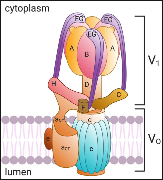
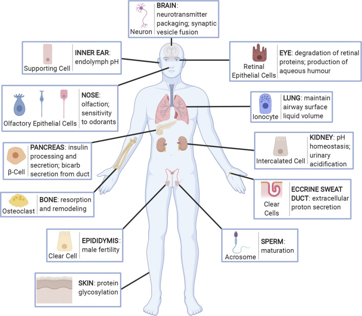
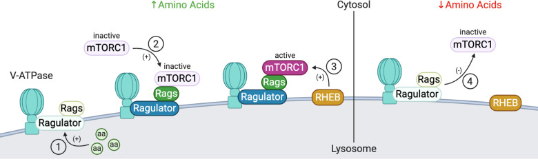
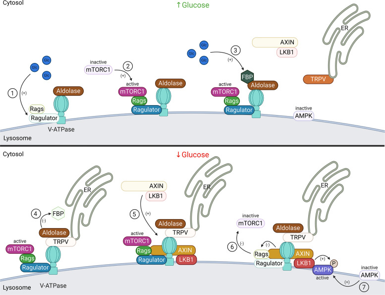
A primary function of the H+-ATPase (or V-ATPase) is to create an electrochemical proton gradient across eukaryotic cell membranes, which energizes fundamental cellular processes. Its activity allows for the acidification of intracellular vesicles and organelles, which is necessary for many essential cell biological events to occur. In addition, many specialized cell types in various organ systems such as the kidney, bone, male reproductive tract, inner ear, olfactory mucosa, and more, use plasma membrane V-ATPases to perform specific activities that depend on extracellular acidification. It is, however, increasingly apparent that V-ATPases are central players in many normal and pathophysiological processes that directly influence human health in many different and sometimes unexpected ways. These include cancer, neurodegenerative diseases, diabetes, and sensory perception, as well as energy and nutrient-sensing functions within cells. This review first covers the well-established role of the V-ATPase as a transmembrane proton pump in the plasma membrane and intracellular vesicles and outlines factors contributing to its physiological regulation in different cell types. This is followed by a discussion of the more recently emerging unconventional roles for the V-ATPase, such as its role as a protein interaction hub involved in cell signaling, and the (patho)physiological implications of these interactions. Finally, the central importance of endosomal acidification and V-ATPase activity on viral infection will be discussed in the context of the current COVID-19 pandemic.

Keywords: acidification; endosomal trafficking; pH regulation; pathophysiology; proton pumping ATPase.


Conflict of interest statement

No conflicts of interest, financial or otherwise, are declared by the authors.

Figures

Figure 1.
Figure 1.
V-ATPase structure and subunit composition. The V-ATPase is composed of a transmembrane (VO) domain comprising the a, c, d, and e subunits and a cytosolic (V1) domain made up of the A, B, C, D, E, F, G, and H subunits. ATP is hydrolyzed at the intersection of the A and B subunits, which powers the rotation of the rotor formed by the d, D, and F subunits. The “c-ring” couples the energy generated by ATP hydrolysis to the translocation of protons from the cytosol to the lumen through the hemichannel formed between the a subunit and the proteolipid c-ring. Created with BioRender.com.
Figure 2.
Figure 2.
Organs and tissues in which V-ATPase membrane expression and proton secretion play a major role in physiological function. Specific V-ATPase holoenzymes are expressed mostly at the apical surface of specialized proton-secreting cells in several tissues throughout the body. These include the supporting cells and interdental cells in the inner ear, epithelial cells in the olfactory mucosa of the nose, β-cells in islets of Langerhans and secretory duct cells in the pancreas, osteoclasts in bone, clear cells in the epididymis, retinal pigment epithelial cells in the eye, ionocytes in the lung, kidney intercalated cells, clear cells in eccrine sweat ducts, and the acrosome of sperm cells. Furthermore, the V-ATPase is expressed ubiquitously in the endomembrane system of cells where it acts to acidify intracellular vesicles. In this capacity the V-ATPase plays particular roles in the brain, for example, where it is involved in the packaging and release of neurotransmitters, as well as soluble N-ethylmaleimide-sensitive factor attachment protein receptor (SNARE)-dependent membrane fusion of synaptic vesicles, and in the skin where it is involved in proper protein glycosylation. Created with BioRender.com.
Figure 3.
Figure 3.
Microscopy of the V-ATPase in proton-secreting cells. The large cytoplasmic V1 domain of the V-ATPase is clearly detectable as an electron dense array of coating material attached to the underside of the lipid bilayer in a kidney intercalated cell apical plasma membrane (A; arrows; bar = 0.1 µm). The rapid-freeze, deep etching procedure reveals the dense, hexagonally packed arrays of V1 domain stud-like projections attached to a vesicle in a proton-secreting cell from toad urinary bladder (B; circles; bar = 25 nm). The freeze fracture technique, exposing the internal domain of split-open lipid bilayers, reveals numerous so-called rod-shaped or dumbbell-shaped (e. g., inside the rectangle) intramembranous particles in V-ATPase-rich membranes: this image is from a kidney intercalated cell (C; bar = 50 nm).
Figure 4.
Figure 4.
Regulation of V-ATPase by vesicle recycling. Recycling of the V-ATPase between intracellular vesicles and the plasma membrane is a mechanism by which proton secretion is modulated. Incorporation of V-ATPase into the apical membrane occurs in response to various physiological stimuli, including several hormones and glucose. One common feature of the process in different cell types is the activation of PKA by increased intracellular cAMP, resulting in phosphorylation (P) of some V-ATPase subunits, including A and C in the cytosolic V1 sector. By an unknown mechanism, this leads to an alteration of the balance between exocytosis (stimulated: green arrow) and endocytosis (reduced: red arrow) of V-ATPase-rich vesicles, resulting in a net accumulation of plasma membrane V-ATPase. One potential stimulus in the kidney and in the epididymis is bicarbonate, generated either by direct entry into the cell through apical bicarbonate exchangers or by the activity of carbonic anhydrase type II in the cytosol. The increased HCO3 concentration stimulates the production of cAMP by soluble adenylyl cyclase (sAC), thereby activating PKA. Phosphorylated V-ATPase complexes are shown at a larger size for clarity. Created with BioRender.com.
Figure 5.
Figure 5.
Different “activation” states of kidney intercalated cells. This plate illustrates regulation of acid secretion by shuttling/recycling of V-ATPase between intracellular vesicles and the plasma membrane. Top: electron micrographs of intercalated cells (IC) from mouse kidney collecting ducts in different states of activation, using immunogold staining with anti-V-ATPase antibodies. The Bottom: schematic representations of the distribution of V-ATPase molecules, vesicles, and apical microvilli that are illustrated at top. A: “nonactivated” cell in which most of the V-ATPase is concentrated in many intracellular vesicles, with very little apical membrane expression (arrow). B: “partially activated” cell in which the V-ATPase is present both in intracellular vesicles, as well as some apical membrane expression coupled with the presence of a few apical microvilli (arrow). C: highly “activated” cell in which most of the V-ATPase is concentrated in extensive apical microvilli and microplicae (arrow), with very little remaining inside the cell on cytoplasmic vesicles. PC, principal cell. Bar = 0.5 µm. (Thank you to Drs. Jennifer Pluznick and Nathan Zaidman, Dept. Physiology, Johns Hopkins Univ. School of Medicine for permission to use EM images prepared as part of an ongoing collaboration.)
Figure 6.
Figure 6.
Effect of CO2 exposure on expression of V-ATPase and apical membrane area of proton-secreting cells in the turtle urinary bladder. Image shows a whole mount view of the apical epithelial surface of a turtle urinary bladder stained with antibodies against the “A” subunit of the V-ATPase, followed by secondary donkey anti-rabbit Ig coupled to Alexafluor 488. The V-ATPase-rich cells (arrows) show a remarkable increase in surface area between the control (A) and tissues acutely exposed to basolateral CO2 (B), indicating that CO2 rapidly activates these cells. Tissue was kindly provided by Dr. John Schwartz, Boston University Medical Center, Boston who exposed bladders to CO2 as part of the annual “Origins of Renal Physiology” course at Mount Desert Island Biological Laboratories, Bar Harbor, ME. Bar = 10 µm.
Figure 7.
Figure 7.
Immunostaining showing type A- and type B-intercalated cells in cortical collecting duct of a rat kidney. Antibodies against the V-ATPase (A-subunit: red), the AE1 anion exchanger (SLC4A1 - yellow), and pendrin (SLC26A4: green) show differential distribution in A- and B-intercalated cells. In this collecting duct, type A cells have apical V-ATPase and basolateral AE1, whereas type B cells have apical pendrin and basolateral V-ATPase. The intensity of basolateral V-ATPase staining in B-cells is lower than the amount of apical staining in the A-cell. Bar = 10 µm. *Tubule lumen.
Figure 8.
Figure 8.
Notch/Wnt signaling pathways. Notch signaling is initiated by ligand binding to Notch receptor (1) leading to receptor endocytosis (2). The receptor is cleaved by γ-secretase in a V-ATPase-dependent manner (3) allowing the cytoplasmic domain to translocate to the nucleus (4) and activate target genes (5). Inhibition of V-ATPase-mediated acidification with bafilomycin blocks this process. Furthermore, V-ATPase-dependent lysosomal degradation of the receptor decreases Notch signaling. Wnt signaling is initiated by Wnt ligand binding to the Frizzled and low-density lipoprotein (LRP) receptors (1) leading to internalization of the receptors into V-ATPase-containing endosomes where they associate with the V-ATPase via the ATP6AP2 accessory protein (2). A multiprotein signaling pathway then inhibits a “destruction complex” that normally degrades β-catenin, leading to an increase in cytoplasmic accumulation and translocation of β-catenin to the nucleus (3), where it activates target genes (4). Created with BioRender.com.
Figure 9.
Figure 9.
V-ATPase-dependent amino acid signaling. Under conditions of high amino acid (aa) availability (left), intralysosomal amino acids activate the Ragulator complex, which acts as a GEF toward the Rag GTPases (Rags), activating them (1). The Rag GTPases are anchored to the lysosomal membrane by the Ragulator complex bound to the V-ATPase. Subsequently, inactive mammalian target of rapamycin complex 1 (mTORC1) is recruited to the lysosomal membrane by the active Rag GTPases (2), where it can be activated by the lysosomal Rheb GTPases (3). Under low amino acid conditions (right), Ragulator GEF activity is inhibited, inactivating Rags, and leading to the disassociation from the lysosome and inactivation of mTORC1 (4). Created with BioRender.com.
Figure 10.
Figure 10.
V-ATPase-dependent glucose signaling. When glucose is present (top), it activates the Ragulator complex, which acts as a GEF toward the Rag GTPases (Rags), activating them (1). The Rag GTPases are anchored to the lysosomal membrane by the Ragulator complex bound to the V-ATPase. Subsequently, mammalian target of rapamycin complex 1 (mTORC1) is recruited to the lysosomal membrane by the active Rag GTPases (2), where it can be activated by the lysosomal Rheb GTPases (see Fig. 9). Additionally, when glucose is present, the glycolytic intermediate fructose-1,6-bisphosphate (FBP) binds to the aldolase-V-ATPase complex (3). Under conditions of glucose starvation (bottom), FBP disassociates from aldolase allowing endoplasmic reticulum (ER)-localized transient receptor potential V-type (TRPV) channels to bind the V-ATPase at the aldolase binding site (4). FBP-unoccupied aldolase binds and inhibits TRPV Ca2+ channel activity allowing for recruitment of the AXIN-LKB1 complex to the lysosomal membrane (5) where it interacts with Ragulator and inhibits its GEF activity toward Rags causing the disassociation from the lysosome and inactivation of mTORC1 (6). Furthermore, the V-ATPase, AXIN, and LKB1 recruit AMPK to form a super complex where AMPK is phosphorylated and activated by LKB1 (7). Created with BioRender.com.

References

    1. Cotter K, Stransky L, McGuire C, Forgac M. Recent insights into the structure, regulation, and function of the V-ATPases. Trends Biochem Sci 40: 611–622, 2015. doi:10.1016/j.tibs.2015.08.005. - DOI - PMC - PubMed
    1. Harrison MA, Muench SP. The facuolar ATPase-A nano-scale motor that drives cell biology. Subcell Biochem 87: 409–459, 2018. doi:10.1007/978-981-10-7757-9_14. - DOI - PubMed
    1. Marshansky V, Rubinstein JL, Gruber G. Eukaryotic V-ATPase: novel structural findings and functional insights. Biochim Biophys Acta 1837: 857–879, 2014. doi:10.1016/j.bbabio.2014.01.018. - DOI - PubMed
    1. Maxson ME, Grinstein S. The vacuolar-type H(+)-ATPase at a glance – more than a proton pump. J Cell Sci 127: 4987–4993, 2014. doi:10.1242/jcs.158550. - DOI - PubMed
    1. Wagner CA, Finberg KE, Breton S, Marshansky V, Brown D, Geibel JP. Renal vacuolar H+-ATPase. Physiol Rev 84: 1263–1314, 2004. doi:10.1152/physrev.00045.2003. - DOI - PubMed

Publication types

MeSH terms

Substances

LinkOut - more resources