Review
. 2021 Mar;115(3):436-452.
doi: 10.1111/mmi.14670. Epub 2021 Jan 7.

Type IV secretion systems: Advances in structure, function, and activation

Affiliations
Review

Type IV secretion systems: Advances in structure, function, and activation

Tiago R D Costa et al. Mol Microbiol. 2021 Mar.

Abstract

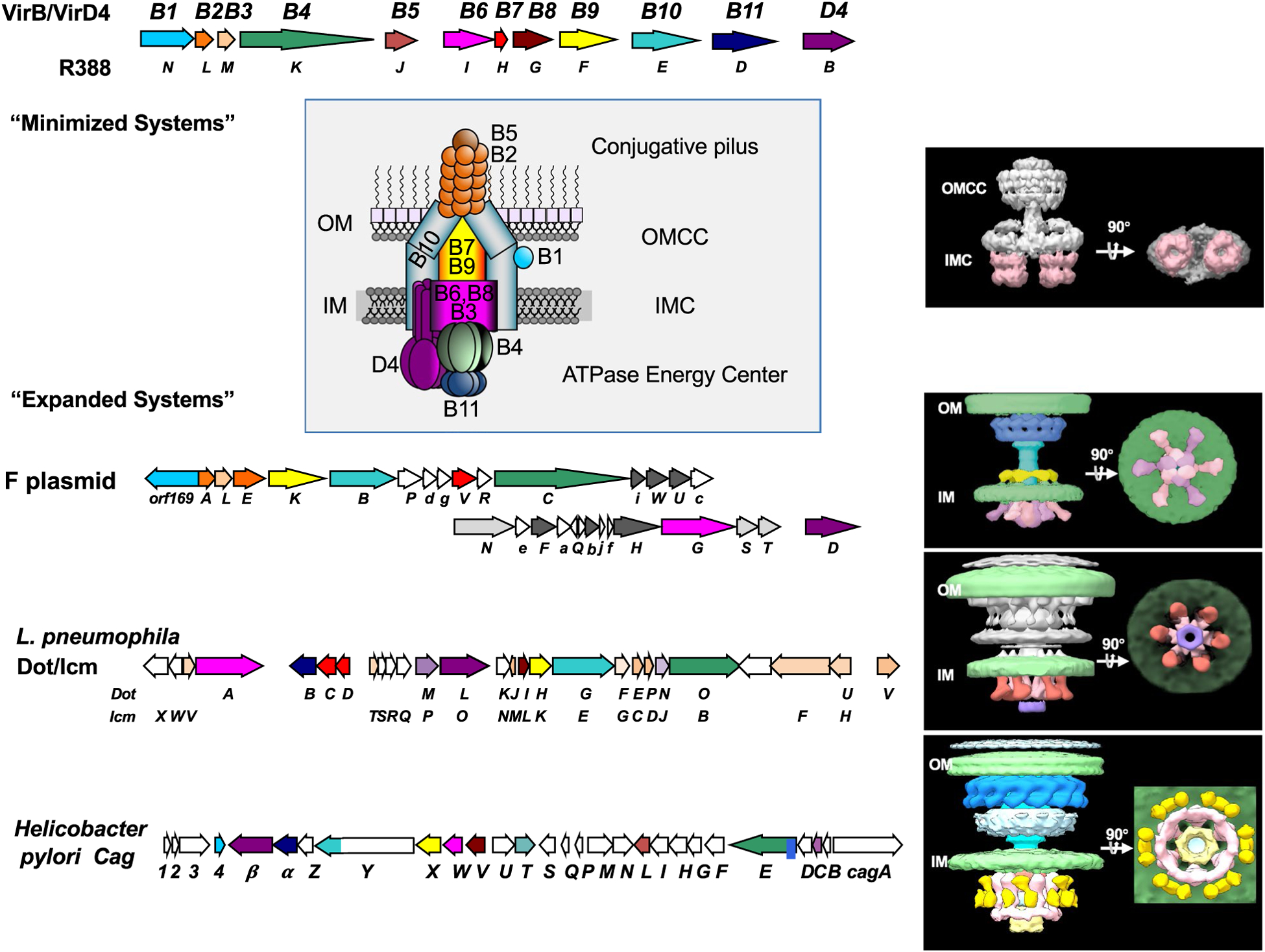
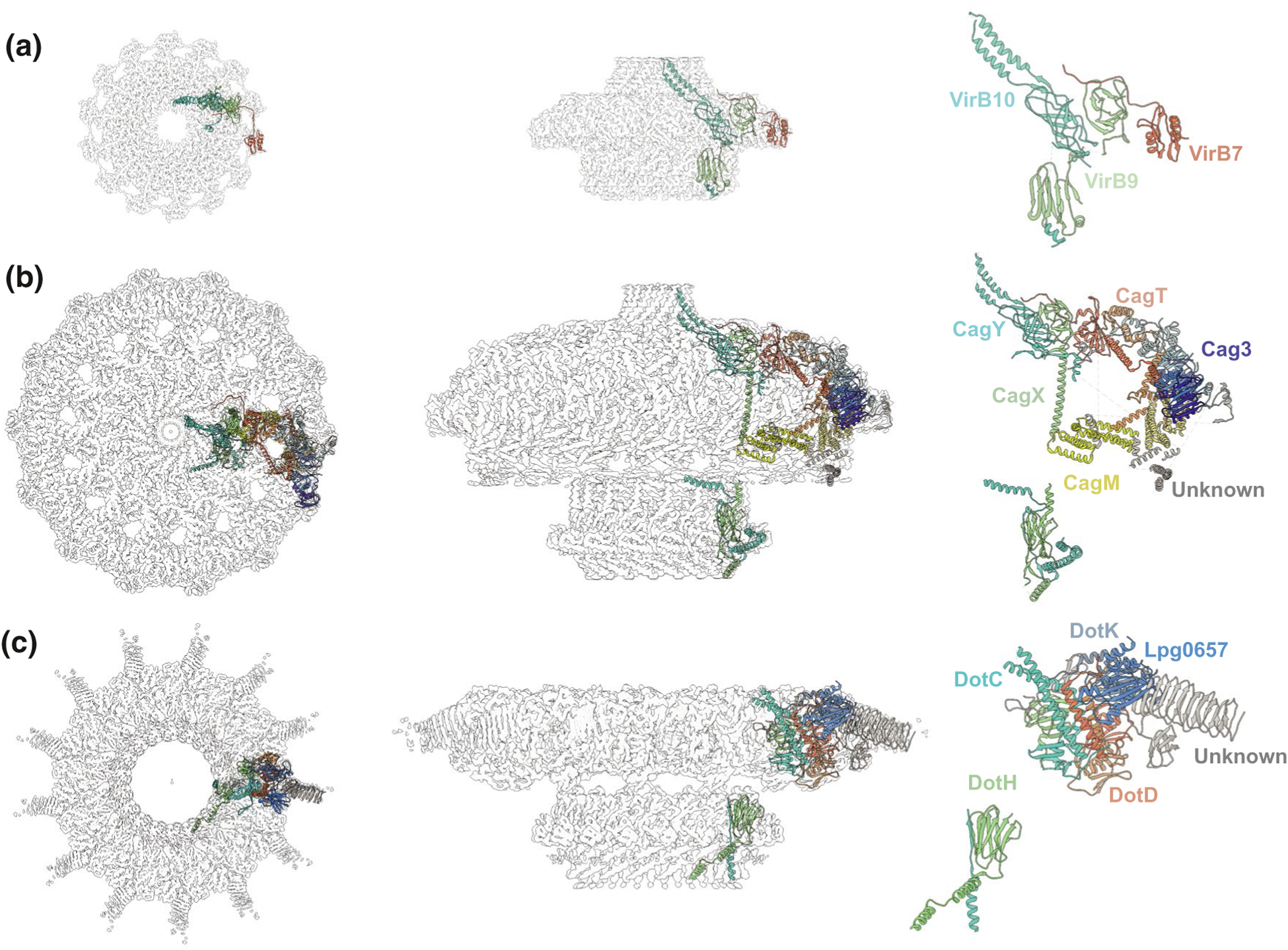
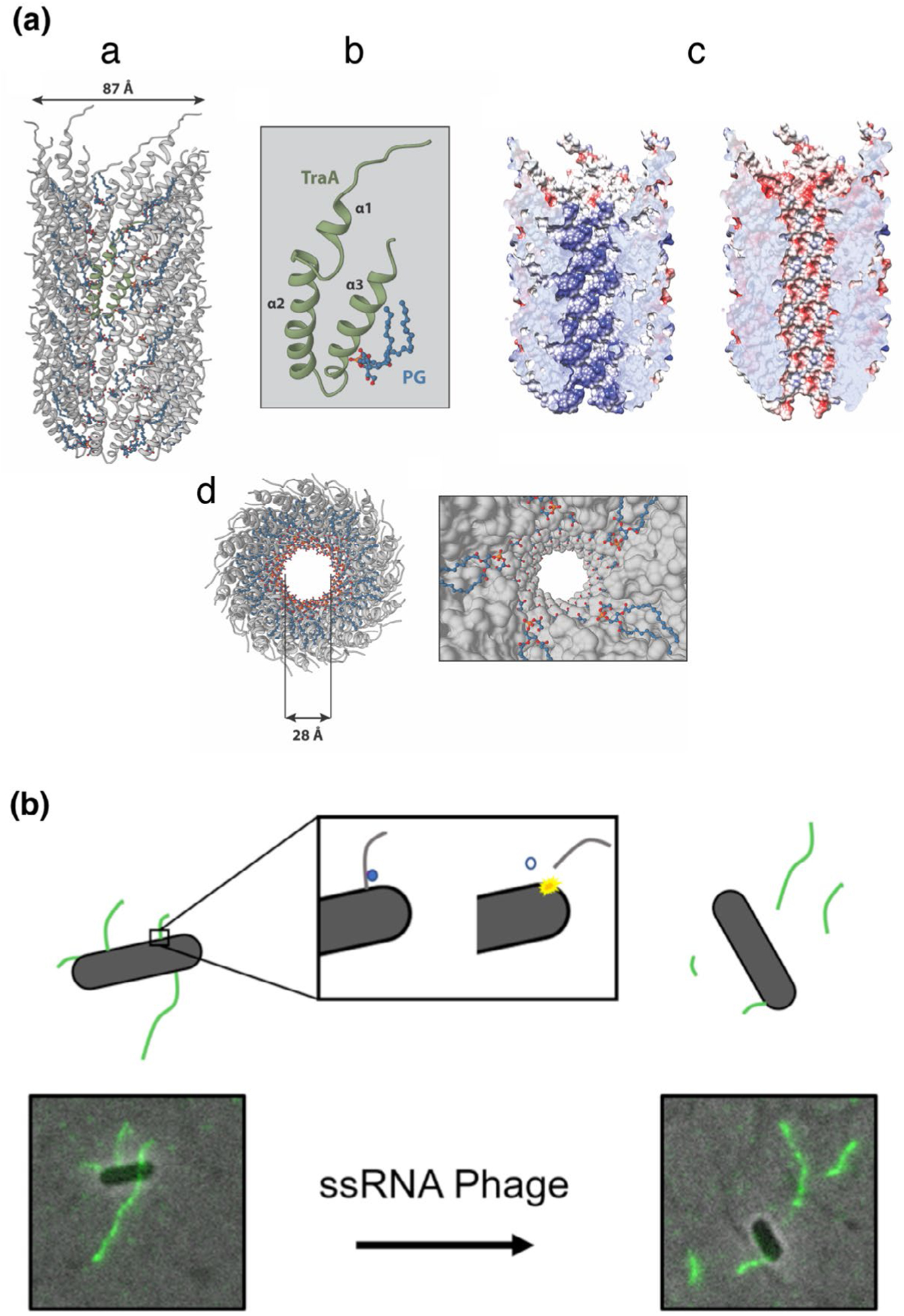
Bacterial type IV secretion systems (T4SSs) are a functionally diverse translocation superfamily. They consist mainly of two large subfamilies: (i) conjugation systems that mediate interbacterial DNA transfer and (ii) effector translocators that deliver effector macromolecules into prokaryotic or eukaryotic cells. A few other T4SSs export DNA or proteins to the milieu, or import exogenous DNA. The T4SSs are defined by 6 or 12 conserved "core" subunits that respectively elaborate "minimized" systems in Gram-positive or -negative bacteria. However, many "expanded" T4SSs are built from "core" subunits plus numerous others that are system-specific, which presumptively broadens functional capabilities. Recently, there has been exciting progress in defining T4SS assembly pathways and architectures using a combination of fluorescence and cryoelectron microscopy. This review will highlight advances in our knowledge of structure-function relationships for model Gram-negative bacterial T4SSs, including "minimized" systems resembling the Agrobacterium tumefaciens VirB/VirD4 T4SS and "expanded" systems represented by the Helicobacter pylori Cag, Legionella pneumophila Dot/Icm, and F plasmid-encoded Tra T4SSs. Detailed studies of these model systems are generating new insights, some at atomic resolution, to long-standing questions concerning mechanisms of substrate recruitment, T4SS channel architecture, conjugative pilus assembly, and machine adaptations contributing to T4SS functional versatility.

Keywords: conjugation; cryoelectron microscopy; cryoelectron tomography; effector translocation; pilus.


Figures

FIGURE 1
FIGURE 1
Structures of T4SSs solved to date. Upper: Operon arrangements of the A. tumefaciens VirB/VirD4 and R388 plasmid-encoded T4SSs, two representative “minimized” systems functioning in Gram-negative species. These systems are assembled from T4SS signature subunits including the 11 VirB subunits and the VirD4 receptor or type IV coupling protein (T4CP). The cartoon depicts locations of the outer membrane core complex (OMCC) and inner membrane complex (IMC) with the associated VirB/VirD4 subunits indicated. Right: Corresponding 3D reconstruction with side and bottom views at 90° angles of the R388-encoded substructure composed of the VirB3 ‒ VirB10 subunits. The substructure was visualized by single-particle negative-stain electron microscopy (EMD-2567); the two side-by-side hexameric barrels of the VirB4 ATPase are highlighted (pink shading). Lower: Operon arrangements of “expanded” systems represented by the F plasmid-encoded Tra, L. pneumophila Dot/Icm, and H. pylori Cag T4SSs. These systems are assembled from the VirB/VirD4 signature subunits (color-coded) plus system-specific subunits (no or gray shading). Right: The F plasmid-encoded Tra (F1-Channel complex, EMD-9344, EMD-9347), L. pneumophila Dot/Icm (EMD-7611, EMD-7612), and H. pylori Cag (EMD-0634, EMD-0635) structures as visualized in the bacterial cell envelope by in situ CryoET. In each of these T4SSs, the VirB4 ATPase presents as a central hexamer of dimers (pink/red-shaded). In the Dot/Icm and Cag machine, the VirB11 homologs DotB (purple-shaded) and Cagα (light yellow shading) dock at the base of the VirB4 homologs DotL and CagE, respectively. In the Cag machine, VirD4-like Cagβ contributes to peripheral densities (yellow and pink)
FIGURE 2
FIGURE 2
High-resolution CryoEM structures from different T4SS outer membrane complexes. (a) The X. citri CryoEM density map (EMDB-0089) resolved at 3.28 Å with 14-fold symmetry; the map is fitted with an asymmetric unit from PDB model 6gyb. (b) The H. pylori CryoEM density maps of the OMC and PR subdomains (OMC:EMD-22081 and PR:20021) resolved at 3.4 Å with 14-fold symmetry and 17-fold symmetry, respectively; the maps are fitted with asymmetric units from PDB models 6×6j and 6×6s. C) The L. pneumophila OMC disk (EMD-22068) resolved at 3.5 Å with 13-fold symmetry and the PR (EMD-22069) resolved at 3.7 Å with 18-fold symmetry, the maps are fitted with asymmetric units from PDB models 6×62 and 6×64. Locations of confirmed subunits are shown, with similar color-coding for the VirB7, VirB9, and VirB10 homologs or orthologs in each system
FIGURE 3
FIGURE 3
F pilus structure and dynamics. A) Architecture of the FpED208 pilus. (a) Representation of the overall molecular model from the FpED208 pilus structure (PDB:5LEG) derived from its cryoelectron density map (EMDB:4042). (b) Molecular model of the F pilus subunit formed by a complex of TraA and PG (phosphatidylglycerol). (c) The electrostatic potential in the pilus lumen in the absence (left) or presence (right) of PG. The blue color represents an electron positive surface, whereas red color represents electron negative. d) Inset view of the pilus lumen showing the PG head group array pointing to the central channel. (b) ssRNA phage penetration causes F pilus detachment. sfGFP fused to the MS2 Coat protein allows for visualization of F pili by proxy of phage binding. Prior to phage infection, F pili are observed protruding from the cell surface. However, once phage is added under infectious conditions, F pili rapidly detach from the cell surface in a time frame matching the MS2 RNA entry period, suggesting that viral RNA penetration triggers detachment of the F pilus

References

    1. Alperi A, Larrea D, Fernandez-Gonzalez E, Dehio C, Zechner EL and Llosa M (2013) A translocation motif in relaxase TrwC specifically affects recruitment by its conjugative type IV secretion system. Journal of Bacteriology, 195, 4999–5006. 10.1128/JB.00367-13 - DOI - PMC - PubMed
    1. Alvarez-Martinez CE and Christie PJ (2009) Biological diversity of prokaryotic type IV secretion systems. Microbiology and Molecular Biology Reviews, 73, 775–808. 10.1128/MMBR.00023-09 - DOI - PMC - PubMed
    1. Alvarez-Rodriguez I, Arana L, Ugarte-Uribe B, Gomez-Rubio E, Martin-Santamaria S, Garbisu C et al. (2020a) Type IV coupling proteins as potential targets to control the dissemination of antibiotic resistance. Frontiers in Molecular Biosciences, 7, 201. 10.3389/fmolb.2020.00201 - DOI - PMC - PubMed
    1. Alvarez-Rodriguez I, Ugarte-Uribe B, de la Arada I, Arrondo JLR, Garbisu C and Alkorta I (2020b) Conjugative coupling proteins and the role of their domains in conjugation, secondary structure and in vivo subcellular location. Frontiers in Molecular Biosciences, 7, 185. 10.3389/fmolb.2020.00185 - DOI - PMC - PubMed
    1. Aly KA and Baron C (2007) The VirB5 protein localizes to the T-pilus tips in Agrobacterium tumefaciens. Microbiology, 153, 3766–3775. 10.1099/mic.0.2007/010462-0 - DOI - PubMed

Publication types

MeSH terms

Substances

LinkOut - more resources