. 2020 Dec 15;33(11):108501.
doi: 10.1016/j.celrep.2020.108501.

CD57+ Memory T Cells Proliferate In Vivo

Affiliations

CD57+ Memory T Cells Proliferate In Vivo

Raya Ahmed et al. Cell Rep. .

Abstract

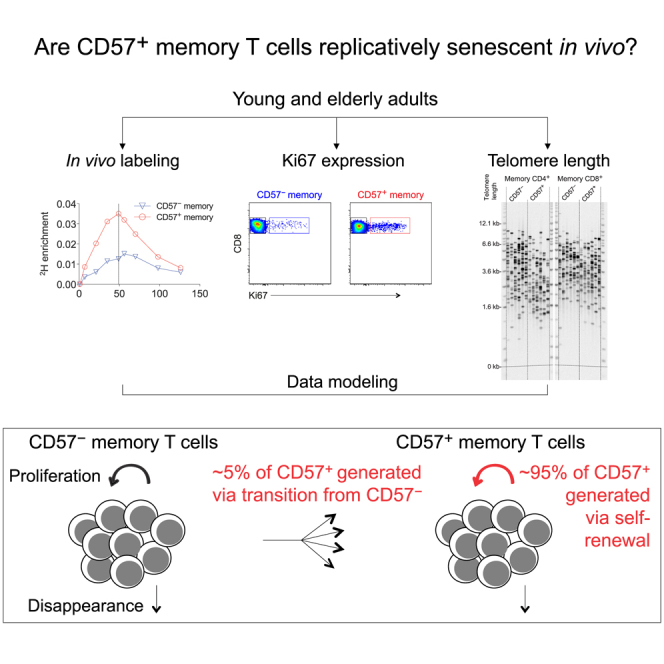
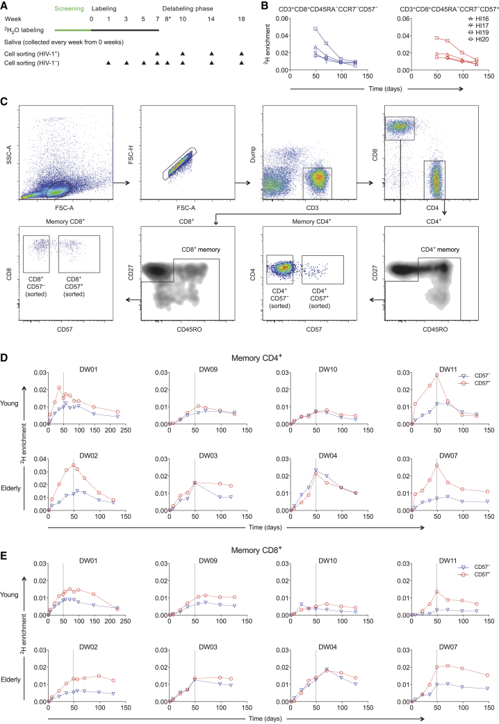
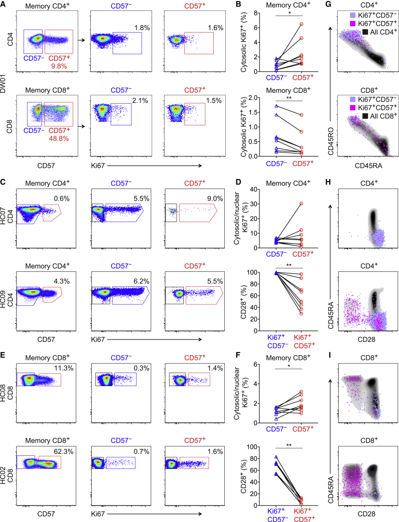
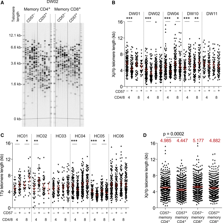
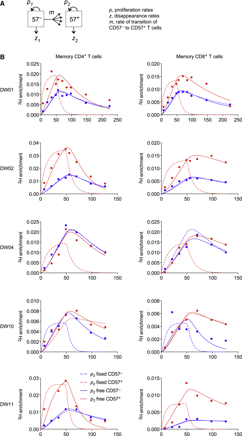
A central paradigm in the field of lymphocyte biology asserts that replicatively senescent memory T cells express the carbohydrate epitope CD57. These cells nonetheless accumulate with age and expand numerically in response to persistent antigenic stimulation. Here, we use in vivo deuterium labeling and ex vivo analyses of telomere length, telomerase activity, and intracellular expression of the cell-cycle marker Ki67 to distinguish between two non-exclusive scenarios: (1) CD57+ memory T cells do not proliferate and instead arise via phenotypic transition from the CD57- memory T cell pool; and/or (2) CD57+ memory T cells self-renew via intracompartmental proliferation. Our results provide compelling evidence in favor of the latter scenario and further suggest in conjunction with mathematical modeling that self-renewal is by far the most abundant source of newly generated CD57+ memory T cells. Immunological memory therefore appears to be intrinsically sustainable among highly differentiated subsets of T cells that express CD57.

Keywords: Enter keywords here.


Conflict of interest statement

Declaration of Interests The authors declare no competing interests.

Figures

None
Graphical abstract
Figure 1
Figure 1
CD57 and CD57+ Memory T Cells Exhibit Similar Rates of Deuterium Incorporation (A) Schematic representation of the 2H2O labeling protocol and sampling time points. (B) Experimental labeling data for CD57 and CD57+ memory CD8+ T cells sampled from the HIV-1-infected volunteers in cohort 1. The corresponding flow cytometric gating strategy is shown in Figure S1. (C) Successive panels depict the flow cytometric gating strategy used to sort CD57 and CD57+ memory T cells from the CD4+ and CD8+ lineages (cohort 2). Lymphocytes were identified in a forward scatter-area versus side scatter-area plot, and single cells were identified in a forward scatter-area versus forward scatter-height plot. Boolean gates were drawn for analysis only to exclude fluorochrome aggregates. Viable CD3+CD14CD19 cells were then identified in the CD4+ and CD8+ lineages, and sort gates were fixed on CD57 and CD57+ memory cells after exclusion of potentially naive CD27brightCD45RO cells. (D) Experimental labeling data for CD57 and CD57+ memory CD4+ T cells sampled from the healthy volunteers in cohort 2. (E) Experimental labeling data for CD57 and CD57+ memory CD8+ T cells sampled from the healthy volunteers in cohort 2.
Figure 2
Figure 2
Ki67+ Cells Are Readily Detectable in the CD57+ Memory T Cell Pool (A) Representative flow cytometric data from a labeled volunteer (DW01) showing cytosolic expression of Ki67 among memory CD4+ (top) or CD8+ T cells (bottom) gated as CD57 (blue) or CD57+ (red). (B) Percent cytosolic expression of Ki67 among memory CD4+ (top) or CD8+ T cells (bottom) gated as CD57 (blue triangles) or CD57+ (red circles). p < 0.05, ∗∗p < 0.01. Paired samples Wilcoxon test. (C) Representative flow cytometric data from unlabeled volunteers (n = 2) showing cytosolic/nuclear expression of Ki67 among memory CD4+ T cells gated as CD57 (blue) or CD57+ (red). HC07 was seronegative for CMV. (D) Top: percent cytosolic/nuclear expression of Ki67 among memory CD4+ T cells gated as CD57 (blue triangles) or CD57+ (red circles). Bottom: percent expression of CD28 among the corresponding Ki67+CD57 (blue triangles) and Ki67+CD57+ memory CD4+ T cells (red circles). ∗∗p < 0.01. Paired samples Wilcoxon test. (E) Representative flow cytometric data from unlabeled volunteers (n = 2) showing cytosolic/nuclear expression of Ki67 among memory CD8+ T cells gated as CD57 (blue) or CD57+ (red). HC02 was seropositive for CMV, and HC08 was seronegative for CMV. (F) Top: percent cytosolic/nuclear expression of Ki67 among memory CD8+ T cells gated as CD57 (blue triangles) or CD57+ (red circles). Bottom: percent expression of CD28 among the corresponding Ki67+CD57 (blue triangles) and Ki67+CD57+ memory CD8+ T cells (red circles). p < 0.05, ∗∗p < 0.01. Paired samples Wilcoxon test. (G) Phenotypic characteristics of Ki67+CD57 and Ki67+CD57+ memory CD4+ (top) or CD8+ T cells (bottom) shown overlaid on density clouds representing the corresponding total CD4+ (top) or CD8+ T cell populations (bottom). Related to (A). (H) Phenotypic characteristics of Ki67+CD57 and Ki67+CD57+ memory CD4+ T cells shown overlaid on density clouds representing the corresponding total CD4+ T cell populations. Related to (C). Key as in (G). (I) Phenotypic characteristics of Ki67+CD57 and Ki67+CD57+ memory CD8+ T cells shown overlaid on density clouds representing the corresponding total CD8+ T cell populations. Related to (E). Key as in (G).
Figure 3
Figure 3
CD57 and CD57+ Memory T Cells Have Similar Division Histories (A) Representative single telomere length analysis (STELA) data showing XpYp telomere lengths among CD57 and CD57+ memory CD4+ or CD8+ T cells sampled from a labeled volunteer (DW02). (B) XpYp telomere lengths among CD57 and CD57+ memory CD4+ or CD8+ T cells sampled from labeled volunteers (cohort 2). p < 0.05, ∗∗p < 0.01, ∗∗∗p < 0.001. Mann-Whitney U test. (C) 17p telomere lengths among CD57 and CD57+ memory CD4+ or CD8+ T cells sampled from unlabeled volunteers (cohort 3). p < 0.05, ∗∗p < 0.01, ∗∗∗p < 0.001. Mann-Whitney U test. (D) Pooled XpYp telomere length data for the volunteers shown in (B). Red lines show means with 95% confidence intervals. Mean values are specified above each column. Significance was assessed using the Mann-Whitney U test.
Figure 4
Figure 4
CD57 and CD57+ Memory T Cells Self-Renew In Vivo (A) Schematic representation of the mathematical model. (B) Model fits to the measured data (dots) for CD57 and CD57+ memory CD4+ (left) or CD8+ T cells (right) with p2 constrained to zero (dashed lines) or free (solid lines).

References

    1. Abo T., Balch C.M. A differentiation antigen of human NK and K cells identified by a monoclonal antibody (HNK-1) J. Immunol. 1981;127:1024–1029. - PubMed
    1. Addo M.M., Draenert R., Rathod A., Verrill C.L., Davis B.T., Gandhi R.T., Robbins G.K., Basgoz N.O., Stone D.R., Cohen D.E. Fully differentiated HIV-1 specific CD8+ T effector cells are more frequently detectable in controlled than in progressive HIV-1 infection. PLoS ONE. 2007;2:e321. - PMC - PubMed
    1. Ahmed R., Westera L., Drylewicz J., Elemans M., Zhang Y., Kelly E., Reljic R., Tesselaar K., de Boer R.J., Macallan D.C. Reconciling estimates of cell proliferation from stable isotope labeling experiments. PLoS Comput. Biol. 2015;11:e1004355. - PMC - PubMed
    1. Ahmed R., Roger L., Costa Del Amo P., Miners K.L., Jones R.E., Boelen L., Fali T., Elemans M., Zhang Y., Appay V. Human stem cell-like memory T cells are maintained in a state of dynamic flux. Cell Rep. 2016;17:2811–2818. - PMC - PubMed
    1. Appay V., van Lier R.A., Sallusto F., Roederer M. Phenotype and function of human T lymphocyte subsets: consensus and issues. Cytometry A. 2008;73:975–983. - PubMed

Publication types

LinkOut - more resources