. 2020 Dec 22;33(12):108528.
doi: 10.1016/j.celrep.2020.108528. Epub 2020 Dec 1.

An ACE2 Microbody Containing a Single Immunoglobulin Fc Domain Is a Potent Inhibitor of SARS-CoV-2

Affiliations

An ACE2 Microbody Containing a Single Immunoglobulin Fc Domain Is a Potent Inhibitor of SARS-CoV-2

Takuya Tada et al. Cell Rep. .

Abstract

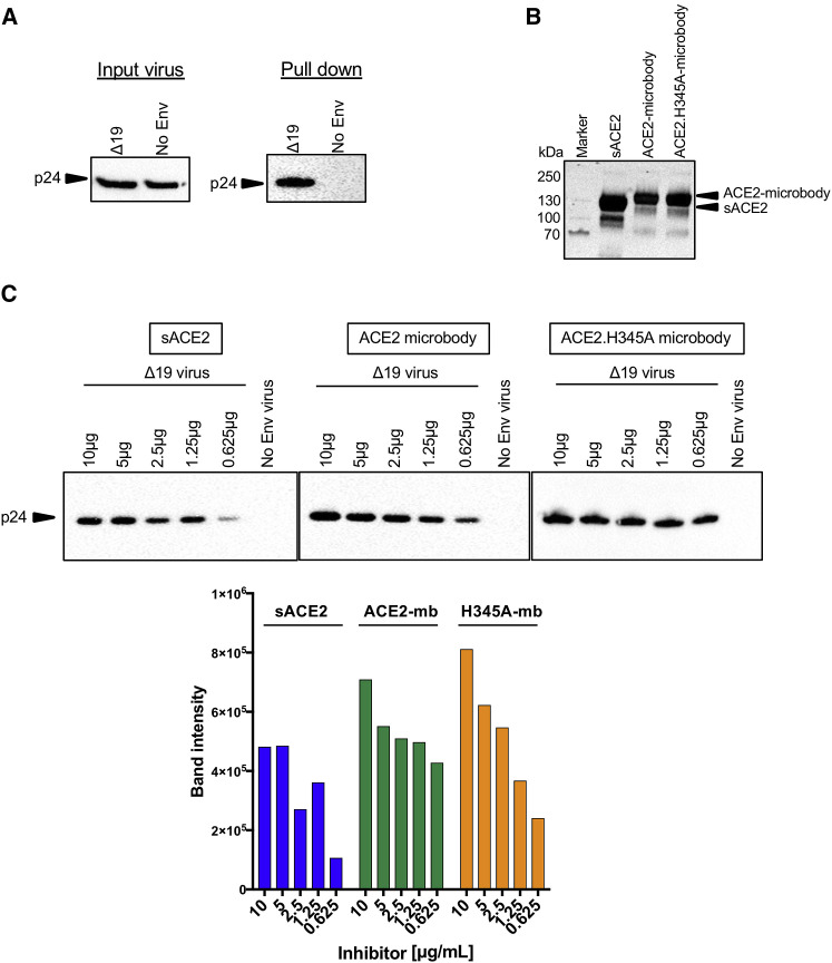
Soluble forms of angiotensin-converting enzyme 2 (ACE2) have recently been shown to inhibit severe acute respiratory syndrome coronavirus 2 (SARS-CoV-2) infection. We report on an improved soluble ACE2, termed a "microbody," in which the ACE2 ectodomain is fused to Fc domain 3 of the immunoglobulin (Ig) heavy chain. The protein is smaller than previously described ACE2-Ig Fc fusion proteins and contains an H345A mutation in the ACE2 catalytic active site that inactivates the enzyme without reducing its affinity for the SARS-CoV-2 spike. The disulfide-bonded ACE2 microbody protein inhibits entry of SARS-CoV-2 spike protein pseudotyped virus and replication of live SARS-CoV-2 in vitro and in a mouse model. Its potency is 10-fold higher than soluble ACE2, and it can act after virus bound to the cell. The microbody inhibits the entry of β coronaviruses and virus with the variant D614G spike. The ACE2 microbody may be a valuable therapeutic for coronavirus disease 2019 (COVID-19) that is active against viral variants and future coronaviruses.

Keywords: ACE2 transgenic; D614G; Fc fusion; SARS-CoV-2; coronavirus; entry inhibitor; lentiviral pseudotype; microbody; soluble ACE2; spike protein.


Conflict of interest statement

Declaration of Interests A provisional patent application has been filed with the US Patent and Trademark Office.

Figures

None
Graphical abstract
Figure 1
Figure 1
Δ19 SARS-CoV-2 Pseudotyped Lentiviral Virion Infection of ACE2.293T Cells and ACE2 Expressing Cell Lines (A) The domain structure of the SARS-CoV-2 spike protein is diagrammed above. Yellow shading indicates the amino acids of the cytoplasmic domain retained following truncation of the 19 carboxy-terminal amino acids (Δ19). Vectors encoding full-length (fl) codon-optimized SARS-CoV-2 spike protein or truncated Δ19 spike protein, with or without a carboxy-terminal HA-tag and the dual nanoluciferase/GFP lentiviral vector pLenti.NLuc.GFP used to generate pseudotyped lentiviral particles, are diagrammed (below). (B) SARS-CoV-2 spike proteins on pseudotyped lentiviral virions were analyzed. Transfected producer cell lysates (left) and supernatant virions (right) were analyzed on an immunoblot probed with anti-HA antibody. Cell lysate blots were probed with anti-GAPDH to normalize protein loading, and virion blots were probed for HIV-1 p24 to normalize for virions. (C) 293T cells transfected with SARS-CoV-2 spike protein expression vectors were analyzed by flow cytometry to detect the protein at the plasma membrane. (D) HA-tagged ACE2 expressed in control transfected 293T and clonal ACE2.293T cells were analyzed on an immunoblot probed with anti-HA antibody. (E) ACE2.293T cells were infected with virus pseudotyped by full-length or SARS-CoV-2 Δ19 spike proteins. Two days post-infection, infectivity was measured by luciferase assay. The reverse transcriptase inhibitor nevirapine was added to one sample to control for free luciferase enzyme contamination of the virus stock. (F) A panel of cell lines were infected with VSV-G, SARS-CoV-2 Δ19 spike protein, or no envelope (no Env) pseudotyped lentivirus (MOI = 0.2). Luciferase activity was measured 2 days post-infection. (G) ACE2.293T cells were treated with ACE2 antibody (1:20) for 30 min at room temperature, and SARS-CoV-2 Δ19 virus was added on the cells. After 2 days of incubation, luciferase activity was measured. The data are represented as the mean of triplicates ± standard deviation. Statistical significance was calculated with the Student’s t test. The experiment was done twice with similar results. IC, intracellular domain; LTR, long terminal repeat; NTD, N-terminal domain; RBD, receptor binding domain; RRE, Rev response element; TM, transmembrane domain.
Figure 2
Figure 2
Wild-Type and H345A ACE2 Microbody Proteins Are Disulfide-Bonded Dimers (A) The domains of ACE2 are shown with the structures of the soluble ACE2 (sACE2), ACE2 microbody, and ACE2.H345A microbody proteins below. The soluble ACE2 proteins are deleted for the transmembrane (TM) and cytoplasmic domains. The ACE2 microbody proteins are fused to the human IgG CH3 domain each with a carboxy-terminal 8× His-tag. (B) The diagram above shows the predicted dimeric structure of the ACE2 microbody protein. The 3D structure of the ACE2:spike protein complex was generated using Chimera software (Pettersen et al., 2004) using published coordinates (Lan et al., 2020). The position of the conserved active site H345 in the ACE2 carboxypeptidase domain is shown lying underneath the ACE2 interaction site. (C) 293T cells were transfected with sACE2, ACE2 microbody, and ACE2.H345A microbody expression vectors. The proteins were pulled down on NTA agarose beads and analyzed under reducing and nonreducing conditions on an immunoblot probed with anti-His-tag antibody. (D) The soluble ACE2 proteins were purified by metal chelate chromatography and size exclusion chromatography (SEC). The oligomerization state was determined by SEC multi-angle light scattering. The calculated molecular mass of each is shown. The experiment was done twice with similar results.
Figure 3
Figure 3
ACE2 Microbody Proteins Bind with High Affinity to SARS-CoV-2 S Pseudotyped Virions Nickel agarose beads were coated for 1 h with 10 μg of soluble ACE2 proteins. Unbound protein was removed, and SARS-CoV-2 Δ19 S pseudotyped virions or virions lacking spike protein were incubated with the beads. After 1 h, unbound virions were removed, and the bound virions were analyzed on an immunoblot probed with anti-p24 antibody. (A) Input virions and bead-bound virions were analyzed on an immunoblot probed with anti-p24 antibody. (B) Soluble ACE2 proteins bound to the nickel agarose beads were analyzed on an immunoblot probed with anti-His-tag antibody. (C) Soluble ACE2, wild-type ACE2 microbody, and ACE2.H345A microbody proteins were serially diluted and bound to nickel agarose beads. The amount of bound virions was determined by immunoblot analysis with anti-p24 antibody. Quantification of the band intensities from the immunoblot is graphed below for soluble ACE2 (sACE2), wild-type ACE2 microbody (ACE2-mb), and ACE2.H345A microbody (H345A-mb). The experiment was done twice with similar results.
Figure 4
Figure 4
ACE2 and ACE2.H345A Microbodies Potently Block Virus Entry and Are Active on Different Cell Lines (A) Serially diluted soluble ACE2, ACE2, and ACE2.H345A microbody proteins were incubated for 30 min with SARS-CoV-2 Δ19.S pseudotyped virus and then added to ACE2.293T cells. Luciferase activity was measured 2 days post-infection. (B) The number of cells infected was determined by flow cytometry to quantify the GFP+ cells. The data are displayed as the percent of GFP+ cells. Representative fluorescence microscopy images of the infected cells are shown below. Scale bar, 50 μm. (C) VSV-G pseudotyped lentiviral virions were incubated for 30 min with 10 μg/mL soluble ACE2 proteins and then added to ACE2.293T cells. Luciferase activity was measured 2 days post-infection. (D) Δ19 spike protein pseudotyped virus was incubated with serially diluted soluble ACE2 proteins for 30 min and then added 293T cells. (E) Serially diluted soluble ACE2 proteins were incubated with mNeonGreen SARS-CoV-2 for 30 min and then added to ACE2.293T cells. After 24 h, the GFP+ cells were counted. Fluorescent microscopy images of representative fields from wells treated with 1 μg soluble ACE2, and ACE2 microbody proteins are shown below. Scale bar, 2.1 mm. The data are displayed as the mean ± SD, and significance is determined by Student’s t tests. The experiments in (A)–(D) were repeated four times, and the experiment in (E) was done twice.
Figure 5
Figure 5
ACE2 Microbody Can Act after Virus/Cell Binding The kinetics of ACE2 microbody inhibition were analyzed in an escape from inhibition assay. (A) The experimental scheme is diagrammed above. SARS-CoV-2 Δ19.S pseudotyped virus was added to ACE2.293T cells. Soluble ACE2 proteins were added immediately or at time points up to 6 h later. Luciferase activity was measured 2 days post-infection. (B) As diagrammed above, virus was bound to target cells for 2 h at 22°C, and unbound virus was then removed. Soluble ACE2 proteins were added as in (A). The data are displayed as the mean ± SD, and statistical significance was determined by Student’s t tests. The experiment was done twice with similar results.
Figure 6
Figure 6
ACE2 Microbody Blocks Entry of the D614G Variant Spike Protein Pseudotyped Virus Infection and ACE2 Using β Coronavirus Spike Proteins (A) The domain structure of the SARS-CoV-2 D614G Δ19 S expression vector is diagrammed above. Red star indicates the D614G mutation in the spike protein. (B) A panel of cell lines was infected with equivalent amounts (MOI = 0.2) of wild-type and D614G Δ19 spike protein pseudotyped virus. (C) Serially diluted soluble ACE2 and ACE2 microbody proteins were mixed with D614G Δ19 spike protein pseudotyped virus and added to target cells. Luciferase activity was measured 2 days post-infection. The data are shown as the mean of triplicates ± SD. The statistical significance of the data was calculated with the Student’s t test. (D) Ni-NTA agarose beads were coated with serially diluted soluble ACE2 and ACE2 microbody proteins. Wild-type, Δ19 spike protein, and no Env pseudotyped virions (28.2 ng p24) were added and allowed to bind. Unbound virions were removed after 30 min, and the bound virions were detected by immunoblot analysis with anti-p24 antibody. The amount (ng) of bead-bound virus p24 was calculated based on band intensities from the immunoblot and indicated in the bottom side. (E) Lentiviral virions pseudotyped by β coronavirus lineage 2 spike proteins were treated with serially diluted soluble ACE2 proteins and then used to infect ACE2.293T cells. The identity of the virus from which the spike protein RBD is derived is indicated above each histogram. Luciferase activity was measured after 2 days. The data are displayed as the mean ± SD, and significance is determined by Student’s t tests. The experiments in (B) and (C) were done three times, and those in (D) and (E) were done twice.
Figure 7
Figure 7
ACE2.H345A Microbody Protects K18-hACE2 Transgenic Mice from SARS-CoV-2 Infection (A and B) ACE2.H345A microbody (7.5 μg) or control buffer was incubated for 30 min at room temperature with viral inoculum (1 × 103 plaque-forming units [PFUs] SARS-CoV-2). The virus was administered intranasally to K18h-ACE2 littermates (control: n = 9; microbody: n = 10), and the mice were then monitored for weight loss (A) and survival (B). Data are pooled from two independent experiments. Weight loss was analyzed by a mixed-effects model. Survival was analyzed by a log rank (Mantel-Cox) test. ∗∗∗∗p < 0.0001.

References

    1. Belouzard S., Millet J.K., Licitra B.N., Whittaker G.R. Mechanisms of coronavirus cell entry mediated by the viral spike protein. Viruses. 2012;4:1011–1033. - PMC - PubMed
    1. Campeau E., Ruhl V.E., Rodier F., Smith C.L., Rahmberg B.L., Fuss J.O., Campisi J., Yaswen P., Cooper P.K., Kaufman P.D. A versatile viral system for expression and depletion of proteins in mammalian cells. PLoS ONE. 2009;4:e6529. - PMC - PubMed
    1. Cantuti-Castelvetri L., Ojha R., Pedro L.D., Djannatian M., Franz J., Kuivanen S., van der Meer F., Kallio K., Kaya T., Anastasina M. Neuropilin-1 facilitates SARS-CoV-2 cell entry and infectivity. Science. 2020;370:856–860. - PMC - PubMed
    1. Carthagena L., Parise M.C., Ringeard M., Chelbi-Alix M.K., Hazan U., Nisole S. Implication of TRIM alpha and TRIMCyp in interferon-induced anti-retroviral restriction activities. Retrovirology. 2008;5:59. - PMC - PubMed
    1. Case J.B., Rothlauf P.W., Chen R.E., Liu Z., Zhao H., Kim A.S., Bloyet L.M., Zeng Q., Tahan S., Droit L. Neutralizing antibody and soluble ACE2 inhibition of a replication-competent VSV-SARS-CoV-2 and a clinical isolate of SARS-CoV-2. bioRxiv. 2020 doi: 10.1101/2020.05.18.102038. - DOI - PMC - PubMed

Publication types

MeSH terms