. 2020 Dec 14;8(12):1991.
doi: 10.3390/microorganisms8121991.

Changes in the Ultrastructure of Staphylococcus aureus Treated with Cationic Peptides and Chlorhexidine

Affiliations

Changes in the Ultrastructure of Staphylococcus aureus Treated with Cationic Peptides and Chlorhexidine

Alina Grigor'eva et al. Microorganisms. .

Abstract

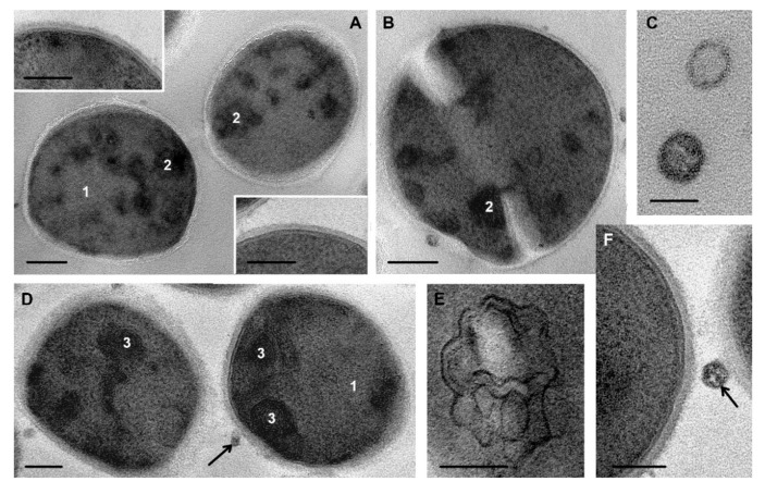
Antimicrobial peptides, including synthetic ones, are becoming increasingly important as a promising tool to fight multidrug-resistant bacteria. We examined the effect of cationic peptides H2N-Arg9-Phe2-C(O)NH2 and H2N-(Lys-Phe-Phe)3-Lys-C(O)NH2 on Staphylococcus aureus, which remains one of the most harmful pathogens. Antiseptic chlorhexidine served as reference preparation. We studied viability of S. aureus and examined its ultrastructure under treatment with 100 µM of R9F2 or (KFF)3K peptides or chlorhexidine using transmission electron microscopy of ultrathin sections. Bacterial cells were sampled as kinetic series starting from 1 min up to 4 h of treatment with preparations. Both peptides caused clearly visible damage of bacteria cell membrane within 1 min. Incubation of S. aureus with R9F2 or (KFF)3K peptides led to cell wall thinning, loss of cytoplasm structure, formation of mesosome-derived multimembrane structures and "decorated fibers" derived from DNA chains. The effect of R9F2 peptides on S. aureus was more severe than the effect of (KFF)3K peptides. Chlorhexidine heavily damaged the bacteria cell wall, in particular in areas of septa formation, while cytoplasm kept its structure within the observation time. Our study showed that cell membrane damage is critical for S. aureus viability; however, we believe that cell wall disorders should also be taken into account when analyzing the effects of the mechanisms of action of antimicrobial peptides (AMPs).

Keywords: S. aureus; cationic peptides; cell membrane; cell wall; chlorhexidine; cytoplasm damage; transmission electron microscopy.


Conflict of interest statement

The authors declare no conflict of interest.

Figures

Figure 1
Figure 1
Viability of S. aureus in the presence of R9F2 and (KFF)3K peptides and chlorhexidine. Viability of S. aureus in 0.9% NaCl solution (saline) and LB broth is presented as a negative control.
Figure 2
Figure 2
S. aureus control (normal) cells. (A) dividing cell; (B) structure of cell periphery; (C) DNA strands (thickness about 3 nm); (D) mesosomes in cytoplasm, note a contact with cell membrane in top cell; (E) cells after division. 1—nucleoid; 2—cytoplasm; 3—cell wall; 4—septum; 5—mesosome. Black arrows show the intermediate layer; white arrows show electron transparent lipid layer of cell membrane, white arrowhead—DNA. TEM of ultrathin sections. Scale bars correspond to: (A)—200 nm, (BE)—100 nm.
Figure 3
Figure 3
Ultrastructure of S. aureus control cells (A); cells after 1 min incubation with R9F2 (B) and (KFF)3K peptides (C,D). 1—cell wall. Black arrows show intermediate layer; white arrows—electron transparent lipid layer of cell membrane. TEM of ultrathin sections. Scale bars correspond to 100 nm.
Figure 4
Figure 4
S. aureus cells: (AC)—control cells; (DF)—cells incubated with peptide R9F2 for 15 min, all cells are deformed and possess “empty” areas in cytoplasm. (GI)—cells incubated with (KFF)3K peptide for 15 min, different degree of damage to cytoplasm is observed. Note the alteration of the intermediate layer and undulation of cell membrane lipid layer on image I. 1—amorphous clumps in cytoplasm; 2—electron dense amorphous cytoplasm at cell periphery; asterisk shows widening of lipid layer. TEM of ultrathin sections. Scale bars correspond to: (A,D,G): 500 nm, other 100 nm.
Figure 5
Figure 5
Ultrastructure of S. aureus cells incubated for 30 min with peptides R9F2 (A,B) and (KFF)3K (C,D). Insert: two cells, demonstrating “normal” cell walls. 1—“empty” areas, 2—areas filled with fine grains; 3—coagulated structureless cytoplasm. Arrow shows mesosome formation. TEM of ultrathin sections. Scale bars correspond to: (A,C)—500 nm, (B,D)—200 nm, inset—100 nm.
Figure 6
Figure 6
Cells of S. aureus incubated for 4 h with R9F2 (A,C) and (KFF)3K (B,D) peptides. 1—multimembrane structures, the insert on image A shows these structure at high magnification, 2—“decorated” fibers, inserts on image C shows “decorated” fibers at high magnification. TEM of ultrathin sections. Scale bars correspond to: (A,C)—200 nm, (B,D)—100 nm, insets—50 nm.
Figure 7
Figure 7
Cells of S. aureus incubated with chlorhexidine for 1 min (A,B) and 15 min (D,F). (C)—membrane vesicles, 1 min oh incubation. (E) mesosome. 1—cytoplasm, 2—electron dense clumps, 3—electron dense masses. Extracellular vesicles are shown with arrows. TEM of ultrathin sections. Scale bars correspond to: (A,B,D,F)—200 nm, (C,E)—100 nm, insets—100 nm.
Figure 8
Figure 8
Multimembrane structures located in areas of septa formation in S. aureus cells incubated with chlorhexidine for 15 min (A); 1 h (B) and 4 h (C). (DF)—enlarged fragments of images (AC). Arrow shows MV. TEM of ultrathin sections. Scale bars correspond to 200 nm.
Figure 9
Figure 9
Cells of S. aureus incubated with chlorhexidine. (A) control cell; (B) 30 min incubation; (C,D) 120 min. Ovals show the areas of septum formation, arrows show the deposition of electron-dense material under cell membrane. TEM of ultrathin sections. Scale bars correspond to 200 nm.
Figure 10
Figure 10
Damage to septa in S. aureus incubated with chlorhexidine. (A) control cells; (B) incubated for 15 min; (C,D) for 45 min; (E) 2 h. Arrows show cell wall in damaged septa. TEM of ultrathin sections. Scale bars correspond to 200 nm.

References

    1. Stryjewski T.P., Zhang F. Effect of Massachusetts health reform on chronic disease outcomes. Health Serv. Res. 2014;49(Suppl. 2):2086–2103. doi: 10.1111/1475-6773.12196. - DOI - PMC - PubMed
    1. Tong S.Y., Davis J.S. Staphylococcus aureus infections: Epidemiology, pathophysiology, clinical manifestations, and management. Clin. Microbiol. Rev. 2015;28:603–661. doi: 10.1128/CMR.00134-14. - DOI - PMC - PubMed
    1. Zouhir A., Jridi T. Inhibition of methicillin-resistant Staphylococcus aureus (MRSA) by antimicrobial peptides (AMPs) and plant essential oils. Pharm. Biol. 2016;54:3136–3150. doi: 10.1080/13880209.2016.1190763. - DOI - PubMed
    1. Kozlova Y.N., Fomenko N.V. Genetic and biochemical characterization of staphylococci occurring in Novosibirsk, Russia. Vavilovskii Zhurnal Genetiki i Selektsii. 2017;21:952–958. doi: 10.18699/VJ17.318. - DOI
    1. Seo M.D., Won H.S. Antimicrobial peptides for therapeutic applications: A review. Molecules. 2012;17:12276–12286. doi: 10.3390/molecules171012276. - DOI - PMC - PubMed

LinkOut - more resources