. 2021 Feb 24;95(6):e00787-20.
doi: 10.1128/JVI.00787-20. Print 2021 Feb 24.

Genome Number and Size Polymorphism in Zika Virus Infectious Units

Affiliations

Genome Number and Size Polymorphism in Zika Virus Infectious Units

Nicole R Sexton et al. J Virol. .

Abstract

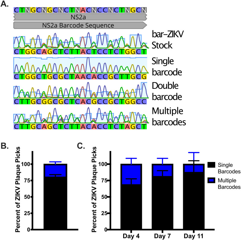
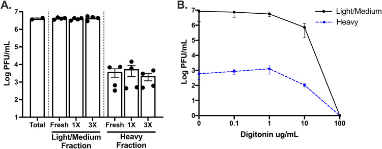
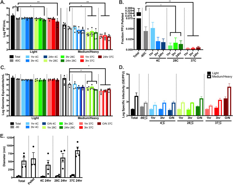
Zika virus (ZIKV; Flaviviridae, Flavivirus) is an arthropod-borne infection that can result in severe outcomes, particularly in fetuses infected in utero It has been assumed that infection by ZIKV, as well as other viruses, is largely initiated by individual virus particles binding to and entering a cell. However, recent studies have demonstrated that multiple virus particles are frequently delivered to a cell simultaneously and that this collective particle delivery enhances infection. ZIKV is maintained in nature between Aedes aegypti mosquitos and vertebrate hosts, including humans. Human infection is initiated through the injection of a relatively small initial inoculum comprised of a genetically complex virus population. Since most mutations decrease virus fitness, collective particle transmission could benefit ZIKV and other arthropod-borne diseases by facilitating the maintenance of genetic complexity and adaptability during infection or through other mechanisms. Therefore, we utilized a barcoded ZIKV to quantify the number of virus genomes that initiate a plaque. We found that individual plaques contain a mean of 10 infecting viral genomes (range, 1 to 212). Few plaques contained more than two dominant genomes. To determine whether multigenome infectious units consist of collectively transmitting virions, infectious units of ZIKV were then separated mechanically by centrifugation, and heavier fractions were found to contain more genomes per plaque-forming unit, with larger diameters. Finally, larger/heavier infectious units reformed after removal. These data suggest that ZIKV populations consist of a variety of infectious unit sizes, likely mostly made up of aggregates, and only rarely begin with a single virus genome.IMPORTANCE The arthropod-borne Zika virus (ZIKV) infects humans and can cause severe neurological sequelae, particularly in fetuses infected in utero How this virus has been able to spread across vast geological ranges and evolve in new host populations is not yet understood. This research demonstrates a novel mechanism of ZIKV transmission through multigenome aggregates, providing insight into ZIKV evolution, immunologic evasion, and better future therapeutic design. This study shows that ZIKV plaques result from collections of genomes rather than individual genomes, increasing the potential for interactions between ZIKV genotypes.

Keywords: ZIKV; Zika virus; aggregates; barcoded virus; flavivirus; specific infectivity.


Figures

FIG 1
FIG 1
Barcode ZIKV contains high diversity at the engineered degenerate sites. (A) ZIKV was engineered with degenerative nucleotides in the third codon position of an 8-amino-acid stretch of NS2a, without altering the protein. (B to D) Barcode diversity is shown as individual barcodes by the percentage of the whole (B), the number of RNA molecules in the sequenced sample for each barcode identified (C), and the percentage of the total stock made up of each barcode (D).
FIG 2
FIG 2
Single and multiple majority barcodes are identified in plaques from bar-ZIKV viral stock, as well as bar-ZIKV-infected mosquito samples. Barcoded ZIKV-infected plaques (Vero cells) were isolated and sent for sequencing across the barcode. (A) Representative chromatograms from plaque purified barcoded ZIKV samples shown. (B and C) Percentages of plaques containing single versus multiple barcode sequences from stock barcoded ZIKV (150 plaques, two independent experiments) (B) or legs/wings from A. aegypti mosquitoes injected with barcoded ZIKV at 4, 7, or 11 dpi presented (47 plaques, two independent experiments) (C). Bars in panels B and C indicate the SD.
FIG 3
FIG 3
The number of genomes contained in a plaque is rarely one. A subset of plaque isolated samples Sanger sequenced were selected to include majority single, double, and multiple barcode-containing plaques. RNA was transcribed to cDNA using PrimerID tags to bioinformatically identify individual RNA molecules. Illumina sequencing was performed across the barcoded region. Individual barcodes are graphed as a percentage of the total barcode population per plaque for single (A), double (B), and multiple (C) majority barcode plaques. (D) The number of unique barcodes per plaque is graphed for each majority group, as is a normalized mean across all plaques. (E) The number of barcodes present at 2.5% of the population or higher is graphed as a function of the total barcodes in each plaque (R2 = Pearson’s correlation coefficient). Barcodes identified were excluded if they were represented by only one RNA molecule, if they contained alternative nucleotides in the first or second codon position within the barcode region, or if they were a minority barcode one mutation away from a well-represented barcode (>50 RNA copies).
FIG 4
FIG 4
ZIKV infectious units can be separated into light and heavy fractions with different particle diameters. Vero cells were infected with a ZIKV infectious clone at an MOI (PFU/cell) of 1 in 10% FBS media (A to C, G and H) or serum-free media (D to F). At 2 dpi (5 dpi for JAR), the supernatants were collected and clarified. (A to D, G and H) Particles in supernatant were separated into three fractions: light (predicted ≤100-nm particle size), medium (predicted 100- to 200-nm particle size), and heavy (predicted ≥200-nm particle size). Washes were performed to remove 50- or 100-nm carryover. Plaque assays were performed to determine fraction titers (A), RT-qPCR from identical samples was performed to determine the genome equivalents (GE) (B), and the specific infectivity was determined (GE/PFU) (C). (D to F) Total or fractionated supernatants were similarly prepared for DLS fixed in 2.5% glutaraldehyde and diluted in PBS. Samples were read on a Zetasizer Nano ZS. (E) Total supernatant from Vero cells is shown as the number of particles in each size group by DLS. (F) DLS measurements were taken for total supernatants generated in JAR (human placental) cells. (G and H) Single- versus multiple-barcode-containing plaques, as determined by using Illumina MiSeq (ultralow variants not present due to a lower read depth). Error bars represent the SEM; comparisons were determined by one-way ANOVA (*, P < 0.05; **, P < 0.005; ***, P < 0.0005; ****, P < 0.0001).
FIG 5
FIG 5
Heavy ZIKV infectious units are not sensitive to freeze-thaw cycles, and the numbers of PFU/ml do not increase after treatment with digitonin. Vero cells were infected with a ZIKV infectious clone at an MOI (PFU/cell) of 1. At 2 dpi, the supernatants were separated into two fractions: light/medium (supernatants from centrifugation at 15,000 × g for 8 min) and heavy (resuspended pellet after centrifugation at 15,000 × g for 8 min). Washes were performed to remove ≤100-nm carryover. Light/medium and heavy fractions were subjected to no, one, or three freeze-thaw cycles (A) or treated with 0, 0.1, 1, 10, or 100 μg/ml of digitonin (B). Plaque assays were performed to determine the fraction titers. Error bars represent the SEM.
FIG 6
FIG 6
Heavy ZIKV infectious units reform after removal, and the particle diameter increases with incubation at increased temperatures. Vero cells were infected with ZIKV at MOI (PFU/cell) of 1 in 10% FBS media (A to C) or serum-free media (D). At 2 dpi, the supernatants were collected and clarified. All particles over a predicted vesicle size of 100 nm were removed by centrifugation at 21,000 × g for 24 min. Supernatants incubated at 4, 28, or 37°C for 1, 3, or 24 h. Immediately after the incubations, the samples were again separated into supernatant (predicted <100 nm) and pellet (predicted >100 nm) fractions, and plaque assays performed to determine the titers (A) and the fractions of PFU pelleted (B). (C) RT-qPCR from identical samples was performed to determine the genome equivalents (GE) used to assess the specific infectivity (GE/PFU). (D) Total or fractionated supernatants were similarly prepared for DLS, followed by fixation in 2.5% glutaraldehyde and dilution in PBS. Samples were read on a Zetasizer Nano ZS. Error bars represent the SEM; comparisons were determined using an unpaired t test (*, P < 0.05; **, P < 0.005).
FIG 7
FIG 7
Replication kinetics are similar between weight separated groups of ZIKV infectious units in different cell types. Vero (A), JAR (B), C636 (C), or Aag2 (D) cells were infected with the titer equilibrized total, using only light or only heavy fractions of ZIKV at an MOI of 0.01 PFU/cell. Supernatant aliquots were taken at indicated days postinfection, and the titers were determined by plaque assay. Error bars represent the SEM (n = 4).

References

    1. Paixão ES, Teixeira MG, Rodrigues LC. 2018. Zika, chikungunya and dengue: the causes and threats of new and re-emerging arboviral diseases. BMJ Glob Health 3:e000530-6. doi: 10.1136/bmjgh-2017-000530. - DOI - PMC - PubMed
    1. Maclachlan NJ, Zientara S, Wilson WC, Richt JA, Savini G. 2019. Bluetongue and epizootic hemorrhagic disease viruses: recent developments with these globally re-emerging arboviral infections of ruminants. Curr Opin Virol 34:56–62. doi: 10.1016/j.coviro.2018.12.005. - DOI - PubMed
    1. Linthicum KJ, Britch SC, Anyamba A. 2016. Rift Valley fever: an emerging mosquito-borne disease. Annu Rev Entomol 61:395–415. doi: 10.1146/annurev-ento-010715-023819. - DOI - PubMed
    1. Leta S, Beyene TJ, De Clercq EM, Amenu K, Kraemer MUG, Revie CW. 2018. Global risk mapping for major diseases transmitted by Aedes aegypti and Aedes albopictus. Int J Infect Dis 67:25–35. doi: 10.1016/j.ijid.2017.11.026. - DOI - PMC - PubMed
    1. Weaver SC, Reisen WK. 2010. Present and future arboviral threats. Antiviral Res 85:328–345. doi: 10.1016/j.antiviral.2009.10.008. - DOI - PMC - PubMed

Publication types