

Two Cases of Post-intubation Laryngotracheal Stenosis Occurring after Severe COVID-19
- PMID: 33328406
- PMCID: PMC7925275
- DOI: 10.2169/internalmedicine.6105-20
Two Cases of Post-intubation Laryngotracheal Stenosis Occurring after Severe COVID-19
Abstract
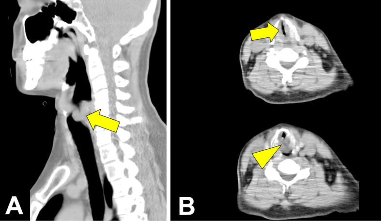
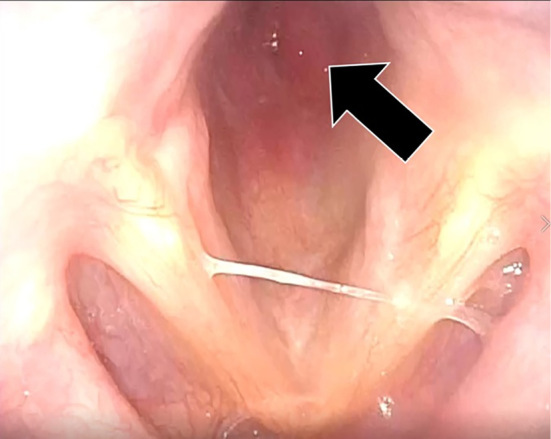
We experienced two cases of post-intubation laryngotracheal stenosis (PILS) occurring in patients after acute coronavirus disease (COVID)-19 in a relatively narrow time period. The patients required mechanical ventilation for 9 days in one and 28 days in the other. In both cases, the patients were discharged but later developed symptoms of cough and dyspnea, which were later diagnosed as PILS. Persistent cough and dyspnea are common symptoms in both PILS and the recovery phase of severe COVID-19. For this reason, PILS should be considered in the differential diagnosis post-COVID-19 patients. In addition, the prevalence of PILS may be greater than that of other critical diseases in severe COVID-19 patients.
Keywords: COVID-19; SARS-CoV-2; coronavirus disease 19; mechanical ventilation; post-intubation laryngotracheal stenosis; tracheal stenosis.
Conflict of interest statement
Figures


References
-
- Farzanegan R, Feizabadi M, Ghorbani F, et al. . An overview of tracheal stenosis research trends and hot topics. Arch Iran Med 9: 598-607, 2017. - PubMed
-
- Nouraei SA, Ma E, Patel A, Howard DJ, Sandhu GS. Estimating the population incidence of adult post-intubation laryngotracheal stenosis. Clin Otolaryngol 5: 411-412, 2007. - PubMed
Publication types
MeSH terms
LinkOut - more resources
Full Text Sources
Other Literature Sources
Medical
Miscellaneous