. 2020 Dec 8:15:9891-9907.
doi: 10.2147/IJN.S283742. eCollection 2020.

Mechanical Properties of Nanohybrid Resin Composites Containing Various Mass Fractions of Modified Zirconia Particles

Affiliations

Mechanical Properties of Nanohybrid Resin Composites Containing Various Mass Fractions of Modified Zirconia Particles

Gaoying Hong et al. Int J Nanomedicine. .

Abstract

Purpose: The aim of this study was to investigate the effect of various mass fractions of 10-methacry-loyloxydecyl dihydrogen phosphate (MDP)-conditioned or unconditioned zirconia nano- or micro-particles with different initiator systems on the mechanical properties of nanohybrid resin composites.

Methods: Both light-cured (L) and dual-cured (D) resin composites were prepared. When the mass fraction of the nano- or micro-zirconia fillers reached 55 wt%, resin composites were equipped with dual-cured initiator systems. We measured the three-point bending-strength, elastic modulus, Weibull modulus and translucency parameter of the nanohybrid resin composites containing various mass fractions of MDP-conditioned or unconditioned zirconia nano- or micro-particles (0%, 5 wt%, 10 wt%, 20 wt%, 30 wt% and 55 wt%). A Cell Counting Kit (CCK)-8 was used to test the cell cytotoxicity of the experimental resin composites. The zirconia nano- or micro-particles with MDP-conditioning or not were characterized by transmission electron microscopy (TEM), Fourier infrared spectroscopy (FTIR), and X-ray photoelectron spectroscopy (XPS).

Results: Resin composites containing 5-20 wt% MDP-conditioned or unconditioned nano-zirconia fillers exhibited better three-point bending-strength than the control group without zirconia fillers. Nano- or micro-zirconia fillers decreased the translucence of the nanohybrid resin composites. According to the cytotoxicity classification, all of the nano- or micro-zirconia fillers containing experimental resin composites were considered to have no significant cell cytotoxicity. The FTIR spectra of the conditioned nano- or micro-fillers showed new absorption bands at 1719 cm-1 and 1637 cm-1, indicating the successful combination of MDP and zirconia particles. The XPS analysis measured Zr-O-P peak area on MDP-conditioned nano- and micro-zirconia fillers at 39.91% and 34.89%, respectively.

Conclusion: Nano-zirconia filler improved the mechanical properties of nanohybrid resin composites, but cannot be the main filler to replace silica filler. The experimental dual-cured composites can be resin cements with better opacity effects and a low viscosity.

Keywords: MDP; initiator system; mechanical property; nano-zirconia filler; resin composite; translucence.


Conflict of interest statement

The authors report no conflicts of interest in this work.

Figures

Figure 1
Figure 1
(A) FTIR spectra of the MDP-conditioned and unconditioned nano-zirconia fillers in the range of 3500–500 cm−1 and peak-fitted FTIR spectra of MDP-conditioned nano-zirconia fillers in the range of 1200–950 cm−1. The peaks corresponding to the P–O bond were located at 985.5 and 1082 cm−1; (B) FTIR spectra of the MDP-conditioned and unconditioned micro-zirconia fillers in the range of 3500–500 cm−1 and peak-fitted FTIR spectra of MDP-conditioned micro-zirconia fillers in the range of 1200–950 cm−1. The peaks corresponding to the P–O bond were located at 972.6 and 1083.7 cm−1. The absorption band of MDP-conditioned nano- and micro-zirconia fillers at 2930 and 2850 cm−1 correspond to the –CH2– stretching vibration. The peaks located at 1719 and 1637 cm−1 were attributed to C=O bond and C=C bond, respectively.
Figure 2
Figure 2
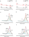
(A) Wide-scan X-ray photoelectron spectroscopy spectra of the unconditioned and MDP-conditioned nano-zirconia fillers; (B) wide-scan X-ray photoelectron spectroscopy spectra of the unconditioned and MDP-conditioned micro-zirconia fillers; (C) narrow-scan O1s spectra of the unconditioned nano-zirconia fillers; (D) narrow-scan O1s spectra of the unconditioned micro-zirconia fillers; (E) narrow-scan O1s spectra of the MDP-conditioned nano-zirconia fillers; and (F) narrow-scan O1s spectra of the MDP-conditioned micro-zirconia fillers. I (C–O), II (P–O–H), III (Zr–O–P), IV (Zr–O–Zr), V (OH–) represent the different deconvoluted peaks within the main peak.
Figure 3
Figure 3
(A) SEM image of the unconditioned micro-silica fillers; (B) SEM image of the unconditioned micro-zirconia fillers; (C) transmission electron micrograph of the unconditioned nano-silica fillers; (D) electron diffraction pattern of the unconditioned nano-silica fillers; (E) transmission electron micrograph of the unconditioned nano-zirconia fillers; and (F) electron diffraction pattern of the unconditioned nano-zirconia fillers.
Figure 4
Figure 4
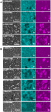
(A) SEM images and EDS maps of light-cured resin composites of L-55si+5zr, L-55si+5Mzr, L-30si+30zr, L-30si+30Mzr; (B) SEM images and EDS maps of dual-cured resin composites of D-55si+5zr, D-55si+5Mzr, D-30si+30zr, D-30si+30Mzr, D-5si+55zr, D-5si+55Mzr. The arrows represent the pits left by the fillers splitting away from the resin surface.
Figure 5
Figure 5
(A) Mean values of three-point bending strength of the light-cured resin composites; (B) mean values of three-point bending strength of the dual-cured resin composites. Same superscript letters (a–d) indicates no significant difference between the groups (P > 0.05).
Figure 6
Figure 6
(A) Weibull distribution plot with 95% confidence intervals of light-cured resin composites; (B) Weibull distribution plot with 95% confidence intervals of dual-cured resin composites.
Figure 7
Figure 7
(A) Mean values of translucency parameter of the light-cured resin composites; (B) mean values of translucency parameter of the dual-cured resin composites. Same superscript letters (a–e) indicates no significant difference between the groups (P > 0.05).
Figure 8
Figure 8
Relative cell proliferations of the experimental groups and the control group.
Figure 9
Figure 9
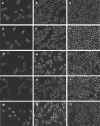
Cell morphology in each group after 1 (A, D, G, J, M), 3 (B, E, H, K, N) and 5 (C, F, I, L, O) days of cell culture. (AC): L-55si+5Zr group; (DF): L-55si+5Mzr group; (GI): D-50si+10zr; (JL): D-50si+10Mzr; and (MO): control group.

References

    1. Broadbent JM, Murray CM, Schwass DR, et al. The dental amalgam phasedown in New Zealand: A 20-year trend. Oper Dent. 2020;45(3):255–264. doi:10.2341/19-024-C - DOI - PubMed
    1. Moraschini V, Fai CK, Alto RM, Santos GOD. Amalgam and resin composite longevity of posterior restorations: A systematic review and meta-analysis. J Dent. 2015;43(9):1043–1050. doi:10.1016/j.jdent.2015.06.005 - DOI - PubMed
    1. Beck F, Lettner S, Graf A, et al. Survival of direct resin restorations in posterior teeth within a 19-year period (1996–2015): A meta-analysis of prospective studies. Dent Mater. 2015;31(8):958–985. doi:10.1016/j.dental.2015.05.004 - DOI - PubMed
    1. Ástvaldsdóttir Á, Dagerhamn J, et al. Longevity of posterior resin composite restorations in adults - A systematic review. J Dent. 2015;43(8):934–954. doi:10.1016/j.jdent.2015.05.001 - DOI - PubMed
    1. Habib E, Wang R, Wang Y, et al. Inorganic fillers for dental resin composites: present and future. ACS Biomater Sci Eng. 2016;2(1):1–11. doi:10.1021/acsbiomaterials.5b00401 - DOI - PubMed