Review
. 2020 Nov 19:11:599729.
doi: 10.3389/fphys.2020.599729. eCollection 2020.

Biological Context Linking Hypertension and Higher Risk for COVID-19 Severity

Affiliations
Review

Biological Context Linking Hypertension and Higher Risk for COVID-19 Severity

Caio A M Tavares et al. Front Physiol. .

Abstract

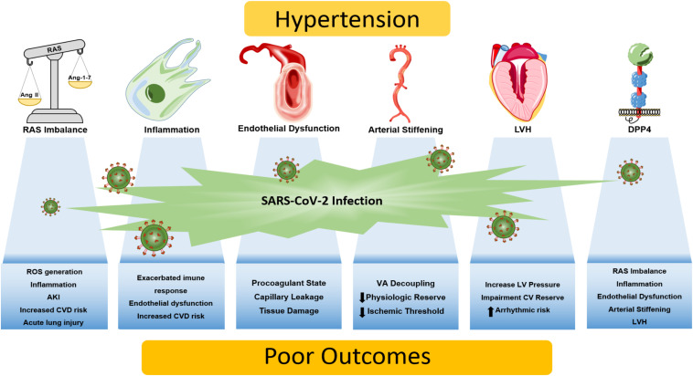
The coronavirus disease 2019 (COVID-19), caused by the severe acute respiratory syndrome coronavirus 2 (SARS-CoV-2), represents a public health crisis of major proportions. Advanced age, male gender, and the presence of comorbidities have emerged as risk factors for severe illness or death from COVID-19 in observation studies. Hypertension is one of the most common comorbidities in patients with COVID-19. Indeed, hypertension has been shown to be associated with increased risk for mortality, acute respiratory distress syndrome, need for intensive care unit admission, and disease progression in COVID-19 patients. However, up to the present time, the precise mechanisms of how hypertension may lead to the more severe manifestations of disease in patients with COVID-19 remains unknown. This review aims to present the biological plausibility linking hypertension and higher risk for COVID-19 severity. Emphasis is given to the role of the renin-angiotensin system and its inhibitors, given the crucial role that this system plays in both viral transmissibility and the pathophysiology of arterial hypertension. We also describe the importance of the immune system, which is dysregulated in hypertension and SARS-CoV-2 infection, and the potential involvement of the multifunctional enzyme dipeptidyl peptidase 4 (DPP4), that, in addition to the angiotensin-converting enzyme 2 (ACE2), may contribute to the SARS-CoV-2 entrance into target cells. The role of hemodynamic changes in hypertension that might aggravate myocardial injury in the setting of COVID-19, including endothelial dysfunction, arterial stiffness, and left ventricle hypertrophy, are also discussed.

Keywords: COVID; dipeptidyl peptidase 4; hemodynamic factors; hypertension; inflammation; renin-angiotensin system.


Figures

FIGURE 1
FIGURE 1
Putative mechanisms linking hypertension and COVID-19 severity. Patients with hypertension are more prone to a vicious interplay between RAS imbalance, chronic low-grade inflammation, and elevated DPP4 activity and expression. Dysregulation of these biological processes may be aggravated by the SARS-CoV-2 infection, giving rise to an exacerbated immune response that culminates in tissue damage/dysfunction. Also, end-organ damage caused by chronic hypertension diminishes cardiovascular reserve, as arterial stiffening, endothelial dysfunction and left ventricular hypertrophy emerges, leading to synergic processes that increase the susceptibility to know complications of COVID-19 including myocardial injury and ischemia, acute lung injury, thrombosis, acute kidney injury, ventricular arrhythmias and potentially death. AKI, acute kidney injury; CV, cardiovascular; CVD, cardiovascular disease; DPP4, dipeptidyl peptidase 4; LV, left ventricle; LVH, left ventricular hypertrophy; RAS, renin-angiotensin system; ROS, reactive oxygen species; VA, ventricle-atrial.

References

    1. Abais-Battad J. M., Lund H., Fehrenbach D. J., Dasinger J. H., Mattson D. L. (2018). Rag1-null Dahl SS rats reveal that adaptive immune mechanisms exacerbate high protein-induced hypertension and renal injury. Am. J. Physiol. Regul. Integr. Comp. Physiol. 315 R28–R35. - PMC - PubMed
    1. Ackermann M., Verleden S. E., Kuehnel M., Haverich A., Welte T., Laenger F., et al. (2020). Pulmonary vascular endothelialitis, thrombosis, and angiogenesis in Covid-19. N. Engl. J. Med. 383 120–128. 10.1056/nejmoa2015432 - DOI - PMC - PubMed
    1. Afify H. M. A., Waits G. S., Ghoneum A. D., Cao X., Li Y., Soliman E. Z. (2018). Peguero electrocardiographic left ventricular hypertrophy criteria and risk of mortality. Front. Cardiovasc. Med. 5:75. 10.3389/fcvm.2018.00075 - DOI - PMC - PubMed
    1. Ainscough J. F., Drinkhill M. J., Sedo A., Turner N. A., Brooke D. A., Balmforth A. J., et al. (2009). Angiotensin II type-1 receptor activation in the adult heart causes blood pressure-independent hypertrophy and cardiac dysfunction. Cardiovasc. Res. 81 592–600. 10.1093/cvr/cvn230 - DOI - PubMed
    1. Aro A. L., Chugh S. S. (2016). Clinical diagnosis of electrical versus anatomic left ventricular hypertrophy: prognostic and therapeutic implications. Circ. Arrhythm. Electrophysiol. 9:e003629. - PMC - PubMed