. 2020 Nov 20:11:593401.
doi: 10.3389/fmicb.2020.593401. eCollection 2020.

The Microbiome of Leonardo da Vinci's Drawings: A Bio-Archive of Their History

Affiliations

The Microbiome of Leonardo da Vinci's Drawings: A Bio-Archive of Their History

Guadalupe Piñar et al. Front Microbiol. .

Abstract

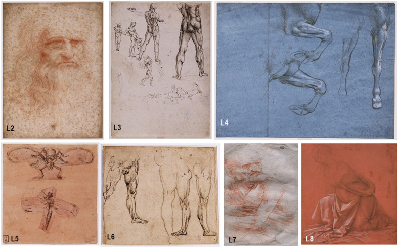
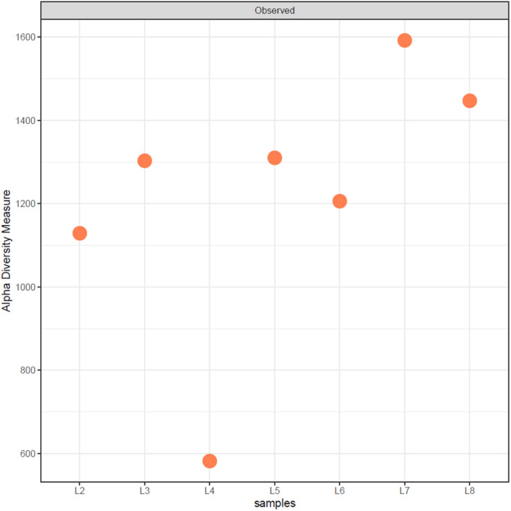
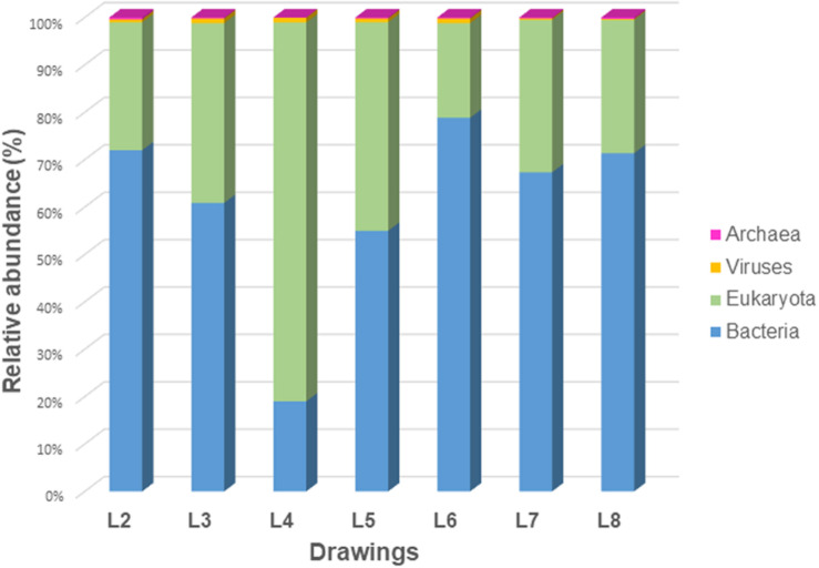
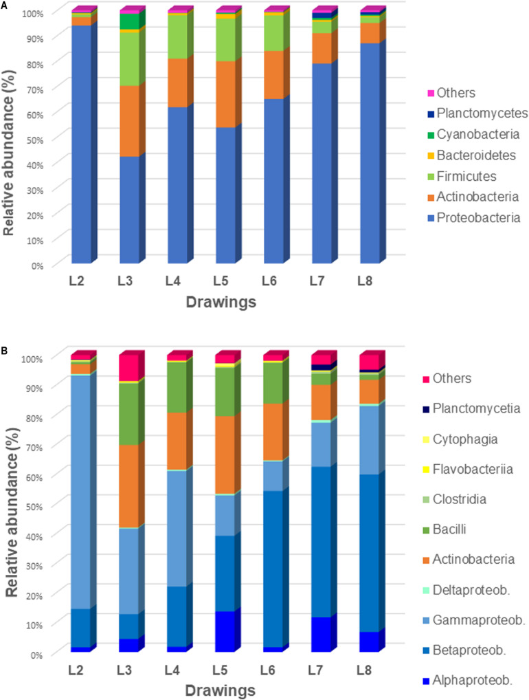
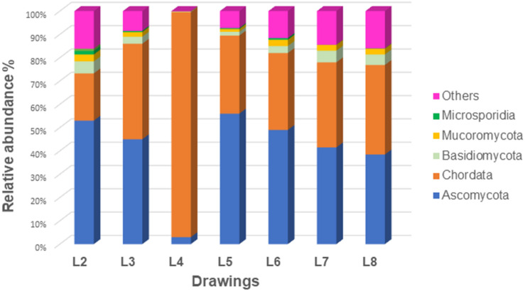
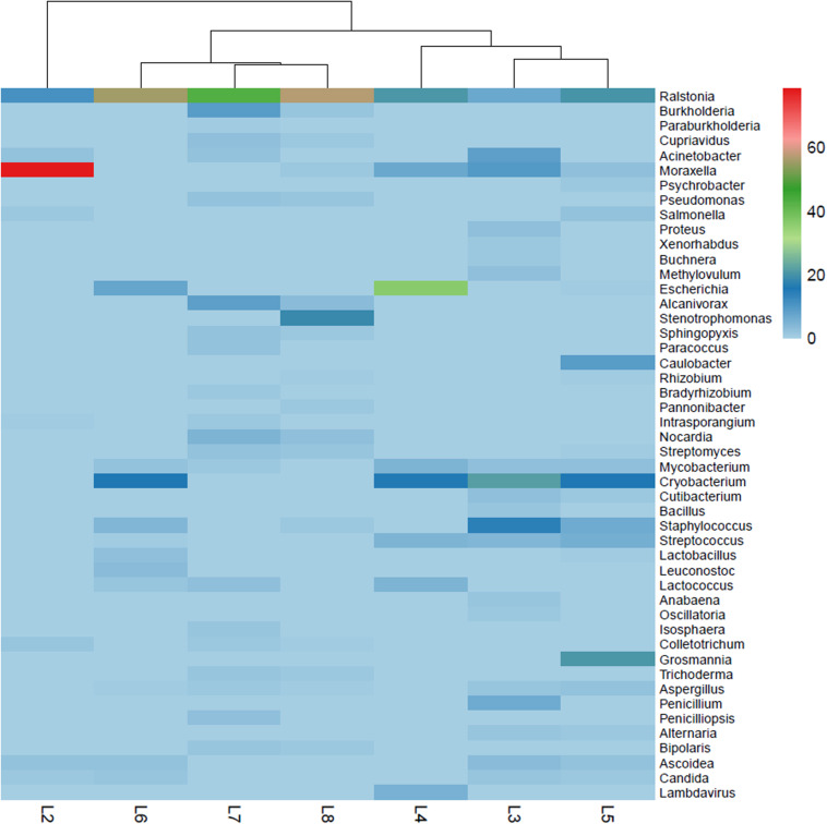
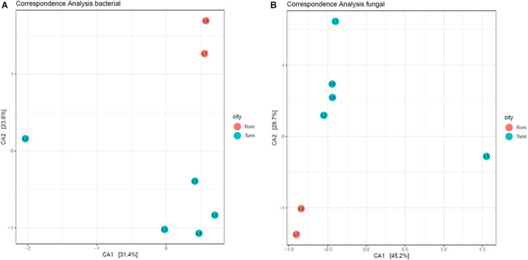
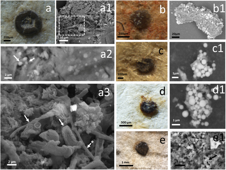
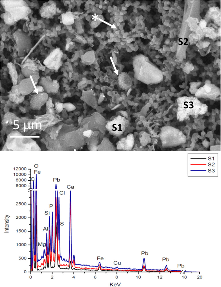
Seven emblematic Leonardo da Vinci's drawings were investigated through third generation sequencing technology (Nanopore). In addition, SEM analyses were carried out to acquire photographic documentation and to infer the nature of the micro-objects removed from the surface of the drawings. The Nanopore generated microbiomes can be used as a "bio-archive" of the drawings, offering a kind of fingerprint for current and future biological comparisons. This information might help to create a biological catalog of the drawings (cataloging), a microbiome-fingerprint for each single analyzed drawing, as a reference dataset for future studies (monitoring) and last but not least a bio-archive of the history of each single object (added value). Results showed a relatively high contamination with human DNA and a surprising dominance of bacteria over fungi. However, it was possible to identify typical bacteria of the human microbiome, which are mere contaminants introduced by handling of the drawings as well as other microorganisms that seem to have been introduced through vectors, such as insects and their droppings, visible through the SEM analyses. All drawings showed very specific bio-archives, but a core microbiome of bacteria and fungi that are repeatedly found in this type of material as true degraders were identified, such as members of the phyla Proteobacteria, Actinobacteria, and Firmicutes among bacteria, and fungi belonging to the classes Sordariomycetes and Eurotiomycetes. In addition, some similarities were observed that could be influenced by their geographical location (Rome or Turin), indicating the influence of this factor and denoting the importance of environmental and storage conditions on the specific microbiomes.

Keywords: Leonardo da Vinci; bio-archive; biological diagnosis; insect droppings; microbiome; nanopore technology; paper material; third generation sequencing.


Figures

FIGURE 1
FIGURE 1
The Leonardo da Vinci’s drawings investigated. The drawings currently housed in the Royal Library of Turin are: “Autoritratto” (sample L2, 33.3 × 21.3 cm), “Nudi per la battaglia di Anghiari” (sample L3, 25.4 × 19.7 cm), “Studi delle gambe anteriori di un cavallo” (sample L4, 15.4 × 20.5 cm), “Studi di insetti” (sample L5, 12.9 × 11.8 cm) and “Studi di gambe virili” recto, “Figura presso il fuoco” verso (sample L6, 14.0 × 15.7 cm). The drawings stored at the Corsinian Library in Rome are: “Uomo della Bitta” (sample L7, 22 × 14.6 cm) and “Studio di panneggio per una figura inginocchiata” (sample L8, 25.7 × 19.0 cm).
FIGURE 2
FIGURE 2
Alpha diversity of drawings calculated from the rarefied classified abundance data using the R package phyloseq.
FIGURE 3
FIGURE 3
Relative abundance of eukaryotes, bacteria, archaea, and viruses in the microbiomes of all seven investigated drawings.
FIGURE 4
FIGURE 4
Relative abundance of the bacterial communities of all seven investigated drawings: (A) at the phylum level and (B) at the class level.
FIGURE 5
FIGURE 5
Relative abundance of the eukaryotic communities of all seven investigated drawings at the phylum level.
FIGURE 6
FIGURE 6
Heatmap reporting the relative abundance (%) of genera in each of the investigated drawings; the genera shown represent greater than 0.5% abundance in at least one sample.
FIGURE 7
FIGURE 7
Canonical correspondence analysis plot of the first and second CA axis for bacterial (A) and fungal (B) abundance data. Dots represent samples (sample name is indicated within the dots) and colors represent the city where the drawings are located—Turin (blue) and Rom (red). Percentages indicate how much of the variability in the data is explained by the respective axis.
FIGURE 8
FIGURE 8
Insects’ droppings. Waxy brown incrustations documented on the fibers of drawing L4 (Studi delle gambe anteriori di un cavallo) and drawing L2 (Autoritratto). These “objects” have been identified as insects’ droppings. They have been documented and, where possible, removed and analyzed by SEM-EDS. (a) an incrustation that was defacing the verso of drawing L4; (a1,a2) SEM images of the waxy material. The images were obtained in variable pressure, with a backscattered detector on non-metallized samples. (a1) shows the appearance of the surface of the object, covered with fungal mycelium, the detail in the square frame corresponds to (a3). The arrows in (a2) show round-shaped crystals embedded in the amorphous matrix. (b,b1,c,c1,d,d1) Correspond to incrustations that were present on the drawing L2. These were made both of an amorphous matrix, as in (b1), but also by spherulites, that consist of spherical crystals made of calcium and magnesium phosphate. (e,e1) Show an incrustation of the L4 drawing where in the amorphous matrix were documented columnar and bipyramidal crystals (arrow).
FIGURE 9
FIGURE 9
SEM images of the surface of a membrane used to sample the surface of the drawing L3 (Nudi per la battaglia di Anghiari). The image was obtained in variable pressure, with a backscattered detector on non-metallized samples. The arrow with the asterisk indicates the appearance of the membrane polymer itself. The other arrows indicate fungal spores (Aspergillus or Penicillium). The EDS spectra corresponding to the labeled particles are reported in the plot below. In this drawing several mineral particles were containing lead, silicon or calcium as the main element.

References

    1. Aich S., Singh R. K., Kundu P., Pandey S. P., Datta S. (2017). Genome-wide characterization of cellulases from the hemi-biotrophic plant pathogen, Bipolaris sorokiniana, reveals the presence of a highly stable GH7 endoglucanase. Biotechnol. Biofuels 10:135. 10.1186/s13068-017-0822-0 - DOI - PMC - PubMed
    1. Alakomi H. L., Paananen A., Suihko M. L., Helander I. M., Saarela M. (2006). Weakening effect of cell permeabilizers on Gram-negative bacteria causing biodeterioration. Appl. Environ. Microbiol. 72 4695–4703. 10.1128/AEM.00142-06 - DOI - PMC - PubMed
    1. Alkhatib N. J., Younis M. H., Alobaidi A. S., Shaath N. M. (2017). An unusual osteomyelitis caused by Moraxella osloensis: a case report. Int. J. Surg. Case Rep. 41 146–149. 10.1016/j.ijscr.2017.10.022 - DOI - PMC - PubMed
    1. Alwadi A., Kilby J., Gawanmeh A. (2017). Tracking and automating a library system using radio frequency identification technology. Int. J. Smart Sens. Intell. Syst. 10 425–450. 10.21307/ijssis-2017-219 - DOI
    1. Andreevskaya M., Johansson P., Laine P., Smolander O. P., Sonck M., Rahkila R., et al. (2015). Genome sequence and transcriptome analysis of meat-spoilage-associated lactic acid bacterium Lactococcus piscium MKFS47. Appl. Environ. Microbiol. 81 3800–3811. 10.1128/AEM.00320-15 - DOI - PMC - PubMed

LinkOut - more resources