. 2020 Nov 20:11:574410.
doi: 10.3389/fimmu.2020.574410. eCollection 2020.

Alpha 1 Antitrypsin-Deficient Macrophages Have Impaired Efferocytosis of Apoptotic Neutrophils

Affiliations

Alpha 1 Antitrypsin-Deficient Macrophages Have Impaired Efferocytosis of Apoptotic Neutrophils

Jungnam Lee et al. Front Immunol. .

Abstract

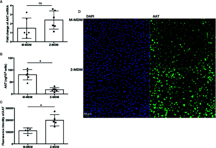
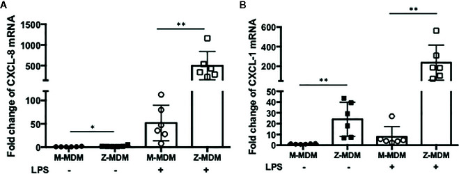
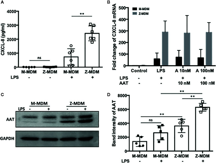
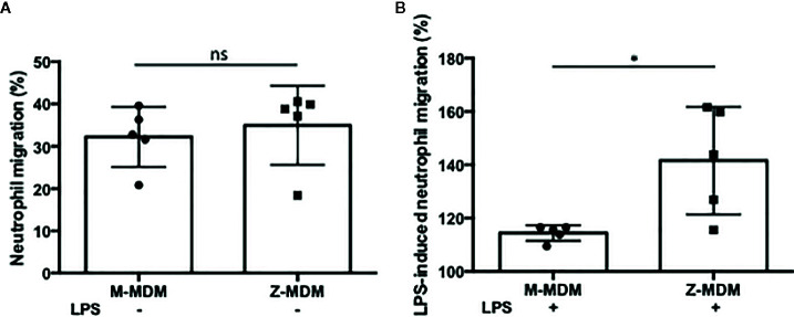
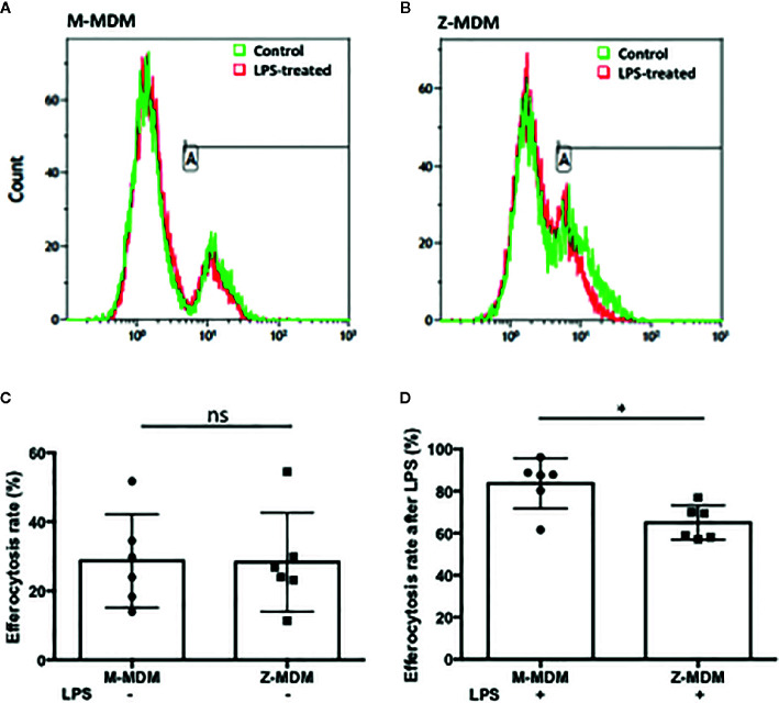
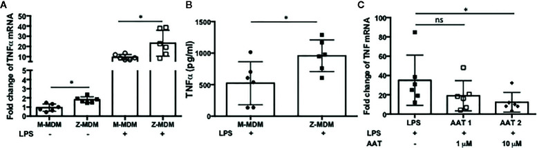
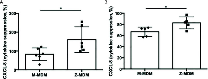
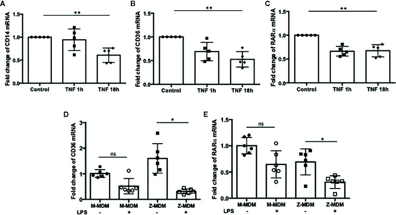
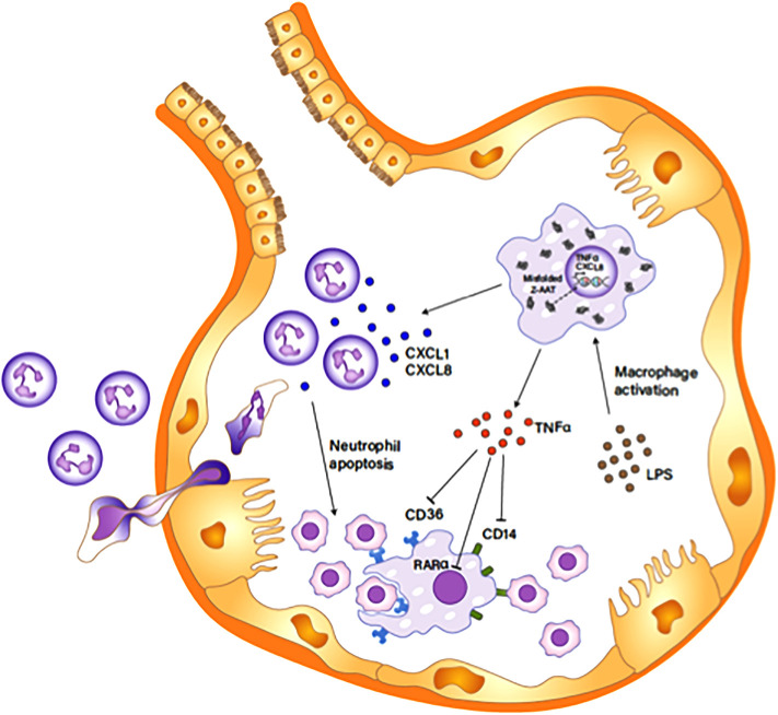
Alpha 1 antitrypsin deficiency (AATD) is an autosomal co-dominant disorder characterized by a low level of circulating AAT, which significantly reduces protection for the lower airways against proteolytic burden caused by neutrophils. Neutrophils, which are terminally differentiated innate immune cells and play a critical role to clear pathogens, accumulate excessively in the lung of AATD individuals. The neutrophil burden in AATD individuals increases the risk for early-onset destructive lung diseases by producing neutrophil products such as reactive oxygen radicals and various proteases. The level of AAT in AATD individuals is not sufficient to inhibit the activity of neutrophil chemotactic factors such as CXCL-8 and LTB4, which could lead to alveolar neutrophil accumulation in AATD individuals. However, as neutrophils have a short lifespan, and apoptotic neutrophils are rapidly cleared by alveolar macrophages that outnumber the apoptotic neutrophils in the pulmonary alveolus, the increased chemotaxis activity does not fully explain the persistent neutrophil accumulation and the resulting chronic inflammation in AATD individuals. Here, we propose that the ability of alveolar macrophages to clear apoptotic neutrophils is impaired in AATD individuals and it could be the main driver to cause neutrophil accumulation in their lung. This study demonstrates that Z-AAT variant significantly increases the expression of pro-inflammatory cytokines including CXCL-8, CXCL1, LTB4, and TNFα in LPS-treated macrophages. These cytokines play a central role in neutrophil recruitment to the lung and in clearance of apoptotic neutrophils by macrophages. Our result shows that LPS treatment significantly reduces the efferocytosis ability of macrophages with the Z-AAT allele by inducing TNFα expression. We incubated monocyte-derived macrophages (MDMs) with apoptotic neutrophils and found that after 3 h of co-incubation, the expression level of CXCL-8 is reduced in M-MDMs but increased in Z-MDMs. This result shows that the expression of inflammatory cytokines could be increased by impaired efferocytosis. It indicates that the efferocytosis ability of macrophages plays an important role in regulating cytokine expression and resolving inflammation. Findings from this study would help us better understand the multifaceted effect of AAT on regulating neutrophil balance in the lung and the underlying mechanisms.

Keywords: AAT deficiency; Alpha 1 antitrysin; cytokine; efferocytosis; macrophage; neutrophil.


Figures

Figure 1
Figure 1
AAT in M- and Z-MDMs. MDMs and their conditioned media were collected at day 7 of macrophage differentiation. AAT mRNA and protein levels are compared in M- and Z-MDMs (n=6). (A) The level of AAT mRNA was measured in M- and Z-MDMs using qRT-PCR. (B) The concentration of AAT was measured in conditioned media of M- and Z-MDMs using ELISA. (C, D) MDMs were immunostained for AAT (green) and the level of intracellular AAT was estimated based on the intensity of positive signal. 20x Images were taken using a fluorescence microscope; bar 50 μm. ~2,000 cells, originating from three separate experiments, were evaluated for each MDM group. *Denotes statistical significance (p-value < 0.05) according to two-tailed Student’s t-test.
Figure 2
Figure 2
LPS-induced neutrophil chemoattractant expression in M- and Z-MDMs. MDMs were incubated with LPS (10 ng/ml) overnight and the expression levels of (A) CXCL-8 and (B) CXCL-1 were compared between M- and Z-MDMs (n=6). The expression levels of the cytokines were normalized to GNB2L1, housekeeping gene.* and **Denote statistical significance (p-value < 0.05 and p-value < 0.01, respectively) according to two-tailed Student’s t-test.
Figure 3
Figure 3
A high level of CXCL-8 resulted from Z-AAT accumulation. MDMs were incubated with LPS (10 ng/ml) overnight. (A) CXCL-8 protein level was measured and compared between LPS-treated M- and Z-MDMs (n=6). (B) Two different concentrations of AAT were extracellularly added to M- and Z-MDM cultures and the cells were incubated with LPS and AAT overnight. Two-way ANOVA test was used to analyze the effect of genotype and treatment (AAT) on the expression of CXCL8 in the cells (n=5). P-values of interaction, genotype, and treatment were 0.96, 0.0001, and 0.996, respectively. *Denotes statistical significance (p-value < 0.05) according to two-tailed Student’s t-test. (C, D) Total proteins were collected from control and LPS-treated MDMs and the increased intracellular AAT levels in LPS-treated cells were visualized and quantified using western blotting (n=5). One-way ANOVA was used to compare the level of intracellular among the samples, and p-value was less than 0.0001. **Denotes statistical significance (p-value < 0.01) according to one-way ANOVA multiple comparisons.
Figure 4
Figure 4
The effect of AAT on LPS-induced neutrophil chemotaxis. MDMs were incubated with LPS (10 ng/ml) overnight. To examine the ability of AAT to regulate neutrophil transmigration, conditioned media of M- and Z-MDMs were collected and placed in the bottom chamber of transwell system. Freshly isolated neutrophils were placed in the top chamber of the transwell system. The number of neutrophils migrated to the bottom layer were counted after 30 min. (A) The number of neutrophils in the bottom layer was divided by the total number of neutrophils to calculate the migration rate of control MDMs. (B) To calculate the migration rate after LPS treatment, the migration rate of control MDM was set to 100%, and the migration rate of LPS-treated MDM was normalized to control for each individual; n=5. *Denotes statistical significance (p-value < 0.05) according to two-tailed Student’s t-test.
Figure 5
Figure 5
Efferocytosis of apoptotic neutrophils by M- and Z-MDMs. MDMs were incubated with LPS (10 ng/ml) overnight and celltracker red-labeled apoptotic neutrophils were added to the MDM culture. Efferocytosis of the apoptotic cells by MDMs was assessed by flow cytometric analysis (A) M-MDM (B) Z-MDM. In each histogram plot, the green and red graphs indicate control and LPS-treated MDMs, respectively. The first peak of the each graph indicates non-phagocytosing MDM population and the second peak indicates phagocytosing MDM population. (C) The efferocytosis rates of MDM controls were assessed by flow cytometric analysis; n=6. (D) Efferocytosis rates of LPS-treated MDMs are expressed relative to those of MDM controls that are set to 100%; n=6. *Denotes statistical significance (p-value < 0.05) according to two-tailed Student’s t-test.
Figure 6
Figure 6
LPS-induced TNFα expression in M- and Z-MDMs. MDMs were incubated with LPS (10 ng/ml) overnight and the gene expression level of (A) TNFα and (B) its protein level were compared between M-MDMs and Z-MDMs. (C) Z-MDMs were incubated with LPS (10 ng/ml) and two different concentrations of M-AAT overnight. The effect of M-AAT on the expression of TNFα was examined in the cells; n=6. *Denotes statistical significance (p-value < 0.05) according to two-tailed Student’s t-test.
Figure 7
Figure 7
The effect of TNFα on macrophage efferocytosis and neutrophil chemotaxis. Z-MDMs were incubated with LPS (10 ng/ml) and TNFα neutralizing antibody overnight. (A) The effect of TNFα neutralizing antibody on neutrophil efferocytosis by MDMs was examined (n=4). For each Z-MDM individual sample, the efferocytosis rate of control MDM was set to 100%, and the efferocytosis rates of the other three treatments were normalized to control MDM. (B) The relative expression levels of CXCL-8 in the LPS-treated cells were calculated in comparison to their non-treated controls (n=3). The CXCL-8 expression level of control MDM was set to 1, and the CXCL-8 expression levels of the other three treatments were normalized to control MDM. (C) Conditioned media of Z-MDM samples were collected for neutrophil chemotaxis assay (n=5). The number of migrated neutrophils was counted, and neutrophil migration rate was calculated based on the number. The migration rate of control MDM was set to 100%, and the migration rates of the other three treatments were normalized to control MDM. *Denotes statistical significance (p-value < 0.05) according to two-tailed Student’s t-test.
Figure 8
Figure 8
Cytokine suppression in post-efferocytotic macrophages. LPS (10 ng/ml)-stimulated MDMs were incubated with apoptotic (A) neutrophils (n=6) or (B) Jurkat cells (n=5) for 3 h. Then, the expression level of CXCL-8 was compared between control and post-efferocytotic cells. Data are expressed as mRNA levels relative to control which are set to 100%. *Denotes statistical significance (p-value < 0.05) according to two-tailed Student’s t-test.
Figure 9
Figure 9
The expression of efferocytosis-related genes regulated by TNFα. MDMs were incubated with TNFα (10 ng/ml) for 1 and 18 h, and the expressions of (A) CD14, (B) CD36, and (C) RARα were examined in the cells (n=5). **Denotes statistical significance (p-value < 0.01) according to one-way ANOVA test. MDMs were incubated with LPS (10 ng/ml) overnight and the relative expression levels of (D) CD36, and (E) RARα were calculated in comparison to non-treated controls for M-MDMs and Z-MDMs (n=6). *Denotes statistical significance (p-value < 0.05) according to two-tailed Student’s t-test.
Figure 10
Figure 10
Z-AAT causing excessive neutrophil accumulation in the pulmonary alveolus. Unfolded Z-AAT induces the expressions of CXCL-8 and TNFα in LPS-stimulated macrophages. The increased level of the chemotactic factor accelerates neutrophil infiltration to the pulmonary alveolus. LPS-induced TNFα expression inhibits the expression of CD14, CD36, and RARα that are important in macrophage efferocytosis of apoptotic cells, delaying the clearance of apoptotic neutrophils by macrophages. The impaired clearance of neutrophils aggravates and prolongs the neutrophil influx in the pulmonary alveolus.

References

    1. Korkmaz B, Attucci S, Jourdan ML, Juliano L, Gauthier F. Inhibition of neutrophil elastase by alpha1-protease inhibitor at the surface of human polymorphonuclear neutrophils. J Immunol (2005) 175(5):3329–38. 10.4049/jimmunol.175.5.3329 - DOI - PubMed
    1. van ‘t Wout EF, van Schadewijk A, Savage ND, Stolk J, Hiemstra PS. alpha1-antitrypsin production by proinflammatory and antiinflammatory macrophages and dendritic cells. Am J Respir Cell Mol Biol (2012) 46(5):607–13. 10.1165/rcmb.2011-0231OC - DOI - PubMed
    1. Janciauskiene SM, Bals R, Koczulla R, Vogelmeier C, Kohnlein T, Welte T. The discovery of alpha1-antitrypsin and its role in health and disease. Respir Med (2011) 105(8):1129–39. 10.1016/j.rmed.2011.02.002 - DOI - PubMed
    1. Henao MP, Craig TJ. Understanding alpha-1 antitrypsin deficiency: A review with an allergist’s outlook. Allergy Asthma Proc (2017) 38(2):98–107. 10.2500/aap.2017.38.4027 - DOI - PubMed
    1. Stoller JK, Aboussouan LS. A review of alpha1-antitrypsin deficiency. Am J Respir Crit Care Med (2012) 185(3):246–59. 10.1164/rccm.201108-1428CI - DOI - PubMed

Publication types

MeSH terms