. 2020 Nov 27:11:561687.
doi: 10.3389/fgene.2020.561687. eCollection 2020.

Xeroderma Pigmentosum C (XPC) Mutations in Primary Fibroblasts Impair Base Excision Repair Pathway and Increase Oxidative DNA Damage

Affiliations

Xeroderma Pigmentosum C (XPC) Mutations in Primary Fibroblasts Impair Base Excision Repair Pathway and Increase Oxidative DNA Damage

Nour Fayyad et al. Front Genet. .

Abstract

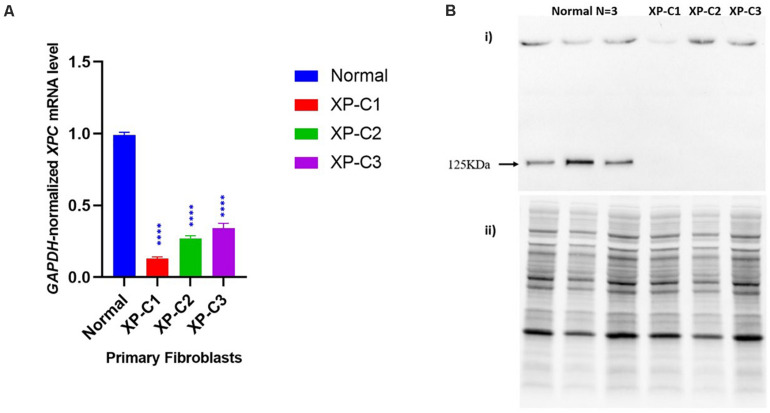
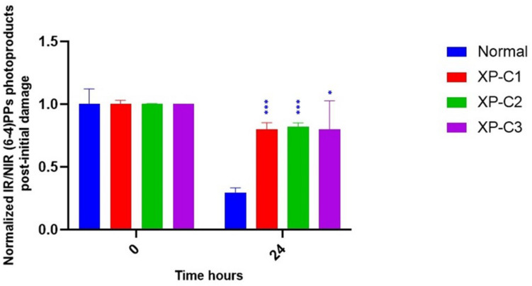
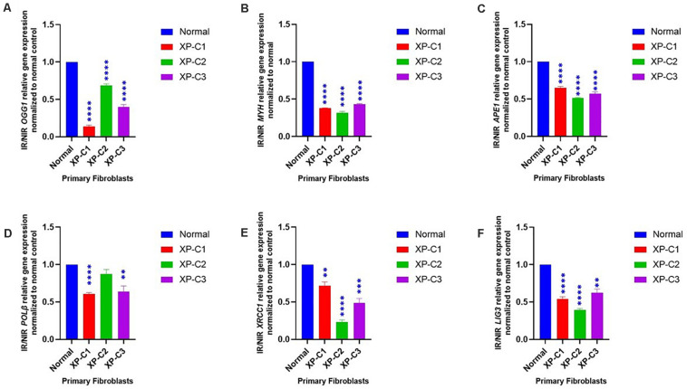
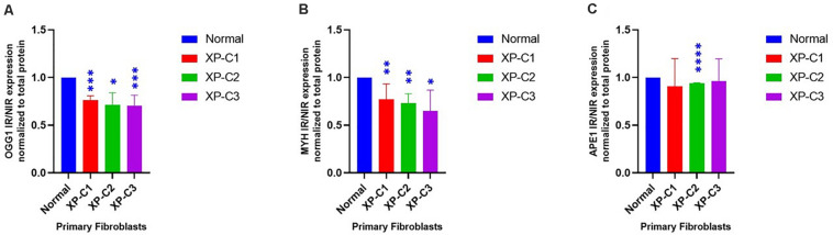
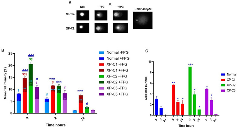
Xeroderma Pigmentosum C (XPC) is a multi-functional protein that is involved not only in the repair of bulky lesions, post-irradiation, via nucleotide excision repair (NER) per se but also in oxidative DNA damage mending. Since base excision repair (BER) is the primary regulator of oxidative DNA damage, we characterized, post-Ultraviolet B-rays (UVB)-irradiation, the detailed effect of three different XPC mutations in primary fibroblasts derived from XP-C patients on mRNA, protein expression and activity of different BER factors. We found that XP-C fibroblasts are characterized by downregulated expression of different BER factors including OGG1, MYH, APE1, LIG3, XRCC1, and Polβ. Such a downregulation was also observed at OGG1, MYH, and APE1 protein levels. This was accompanied with an increase in DNA oxidative lesions, as evidenced by 8-oxoguanine levels, immediately post-UVB-irradiation. Unlike in normal control cells, these oxidative lesions persisted over time in XP-C cells having lower excision repair capacities. Taken together, our results indicated that an impaired BER pathway in XP-C fibroblasts leads to longer persistence and delayed repair of oxidative DNA damage. This might explain the diverse clinical phenotypes in XP-C patients suffering from cancer in both photo-protected and photo-exposed areas. Therapeutic strategies based on reinforcement of BER pathway might therefore represent an innovative path for limiting the drawbacks of NER-based diseases, as in XP-C case.

Keywords: Xeroderma Pigmentosum C; base excision repair; nucleotide excision repair; oxidative DNA damage; oxidative stress; skin cancer; ultra violet (UV) light.


Conflict of interest statement

The authors declare that the research was conducted in the absence of any commercial or financial relationships that could be construed as a potential conflict of interest.

Figures

FIGURE 1
FIGURE 1
Impaired XPC mRNA and protein levels in XP-C primary fibroblasts at basal level. (A) XPC mRNA level is downregulated in XP-C primary fibroblasts compared to normal control at basal level. Upon qRT-PCR, all three XP-C fibroblasts exhibited a significantly reduced XPC gene transcription compared to the normal (n = 3). (p < 0.0001 ****, Unpaired-t-test, GraphPad Prism 8). The data were normalized relative to the GAPDH mRNA levels, where GAPDH was used as an endogenous control. The results are the mean ± SEM from three independent experiments, n = 3. (B) XPC protein was not expressed in XP-C primary fibroblasts at basal level. Although XPC protein was readily observed in all three normal fibroblasts, it was undetectable in the three XP-C fibroblasts at MW = 125 KDa. The results correspond to the mean ± SEM from three independent experiments, n = 3. (i) membrane with XPC bands upon hybridization with anti-XPC (ii) total protein-membrane used for normalization.
FIGURE 2
FIGURE 2
Similar Photosensitivity between normal and XP-C primary fibroblasts. Short-term cytotoxicity test (MTT) was done 24 h post-UVB-irradiation. This was done by comparing the cellular viability between normal and XP-C fibroblasts at each UVB dose condition. Each sample was normalized by its non-irradiated value (100% viability). Unpaired-t-test was used to compare photosensitivity between normal and each XP-C fibroblast at each UVB dose (GraphPad Prism 8). The results are the mean ± SEM from three independent experiments, n = 3.
FIGURE 3
FIGURE 3
Downregulated repair of (6–4) PPs, bulky photoproducts, in XP-C fibroblasts compared to normal control. Immunocytochemistry was done to detect (6–4) PPs by fixation instantaneously at 0 and 24 h post-UVB-irradiation (0.03 J/cm2). An absence of primary antibody was used as negative control. The nuclei were stained with Hoechst and the (6–4) PPs were detected by green fluorescently labeled primary antibody. Images were shown upon merging both fluorescence, thereby, lesions were quantified (fluorescence signal) and normalized by non-irradiated conditions. XP-C1, XP-C2, and XP-C3 showed a significant persistence of lesion repair at 24 h compared to normal (p < 0.001,∗∗∗; p < 0.001,∗∗; p < 0.05, respectively). Unpaired-t-test was used to compare normalized IR/NIR lesion between normal and each XP-C fibroblast at each UVB dose (0 and 24 h) (GraphPad Prism 8). The results are the mean ± SEM from two independent experiments, n = 2 (each experiment is done as a triplicate). IR, irradiated, NIR, non-irradiated.
FIGURE 4
FIGURE 4
Downregulated BER-associated gene transcription in normal and XP-C fibroblasts, post-UVB-irradiation. Gene transcription was investigated by qRT-PCR experiments in XP-C vs. control fibroblasts 4 h post-UVB dose (0.05 J/cm2). Total RNA extraction was followed by reverse transcription. QRT-PCR was carried out to assess gene expression. Shown values are the mean ± SEM from three independent experiments, n = 3. The used calibrator was non-irradiated normal fibroblast where expression ratios were normalized by that of control. Ratio of IR/NIR was used in analysis. Panel (A) shows the significant downregulation of normalized IR/NIR OGG1 gene expression in XP-C fibroblasts compared to normal (p < 0.0001,****). Panel (B) shows the significant downregulation of normalized IR/NIR MYH gene expression in XP-C fibroblasts compared to normal (p < 0.0001,****). Panel (C) shows the significant downregulation of normalized IR/NIR APE1 gene expression in XP-C fibroblasts compared to normal (p < 0.0001,****). Panel (D) XP-C1 and XP-C3 showed a significant Polβ downregulation compared to normal (p < 0.0001,**** and p < 0.01,∗∗ respectively) while no significant difference was observed while comparing XP-C2 to the control. Panel (E) shows the significant downregulation of normalized IR/NIR XRCC1 gene expression in XP-C1, XP-C2, and XP-C3 compared to normal (p < 0.01,∗∗; p < 0.0001,**** and p < 0.001,∗∗∗ respectively). Panel (F) shows the significant downregulation of normalized IR/NIR LIG3 gene expression in XP-C1, XP-C2, and XP-C3 compared to normal (p < 0.0001,****; p < 0.0001,**** and p < 0.01,∗∗ respectively). This was done by unpaired-t-test that allows the comparison between normal and each XP-C fibroblast (GraphPad Prism 8). IR, irradiated; NIR, Non-Irradiated.
FIGURE 5
FIGURE 5
Downregulated BER-associated protein levels in normal and XP-C fibroblasts, post-UVB-irradiation. Protein level was investigated in XP-C vs. control fibroblasts 4 h post-UVB-irradiation (0.05 J/cm2). Total protein was extracted followed by western blot to evaluate protein expression. Values shown are the mean ± SEM from three independent experiments, n = 3. Ratio of IR/NIR was used in analysis after normalization by the total protein. Panel (A) shows the significant downregulation of normalized IR/NIR OGG1 protein expression in XP-C1, XP-C2, and XP-C3 compared to normal (p < 0.001,∗∗∗; p < 0.05,; and p < 0.001,∗∗∗). Panel (B) shows the significant downregulation of normalized IR/NIR MYH protein expression in XP-C1, XP-C2, and XP-C3 compared to normal (p < 0.01,∗∗; p < 0.01,∗∗ and p < 0.05, respectively). Panel (C) shows the significant downregulation of normalized IR/NIR APE1 gene expression in XP-C2 fibroblast compared to normal (p < 0.0001,****). Statistical analysis was done by unpaired-t-test that allows the comparison between normal and each XP-C fibroblast (GraphPad Prism 8). IR, irradiated; NIR, Non-Irradiated.
FIGURE 6
FIGURE 6
Low intrinsic base excision excision-repair capacities in XP-C primary fibroblasts. Comet ± FPG was done to detect single-strand breaks (SSB), alkali-labile sites (ALS), and oxidative purines (including 8-oxoGua) in each sample at each condition. (A) Illustrates the undamaged (comet head) and damaged (comet tail) DNA ± FPG in normal and XP-C1 fibroblasts and positive control H2O2; the% tail intensity and length are proportional to the DNA damage. (B) The graphical representation displays the mean tail intensities (%) for each sample, for both FPG active sites (dark-colored) and SSB/ALS (light-colored) post-UVB-irradiation (0.05 J/cm2). All fibroblasts were able to repair; however, the XP-C fibroblasts had a downregulated and dwindled repair activity. We did a ratio of IR/NIR ± FPG for each fibroblast at three kinetic points = 0, 2, and 24 h. The results are the mean ± SEM from three independent experiments. Paired-t-test was done to compare each sample with two conditions (FPG–ve or FPG+ve). Unpaired-t-test was done to compare different samples within the same condition (GraphPad Prism 8). $ Sample significantly (p < 0.05) higher in its tail intensity with presence of FPG (+FPG) compared to its absence (–FPG). XP-C fibroblast significantly (p < 0.05) higher in its tail intensity compared to normal fibroblast, at –FPG condition. μ XP-C fibroblast significantly (p < 0.05) higher in its tail intensity compared to normal fibroblast, at +FPG condition. (C) The graphical representation displays oxidized purines repair (8-oxoGua and Fapy) in normal compared to XP-C fibroblasts. These oxidized purines were detected upon subtracting values +FPG from values –FPG for each sample. As expected, oxidized purines were downregulated in all fibroblasts. At t = 0 h, XP-C1, XP-C2, and XP-C3 had higher oxidized purines compared to normal fibroblasts (p < 0.01,∗∗; p < 0.001,∗∗∗ and p < 0.05,∗∗ respectively). Similarly was shown at t = 2 and 24 h for XP-C1, XP-C2, and XP-C3; except for XP-C3 at 24 h. Shown values correspond to the mean ± SEM from three independent experiments. Unpaired-t-test was done to compare different samples within the same condition (GraphPad Prism 8). XP-C fibroblast significantly (p < 0.05) higher in its oxidized purines compared to normal fibroblast. –FPG = FPG alkaline buffer without the enzyme. +FPG = FPG alkaline buffer and FPG enzyme. IR, irradiated, NIR, Non-Irradiated.

References

    1. Ba X., Boldogh I. (2018). 8-Oxoguanine DNA glycosylase 1: beyond repair of the oxidatively modified base lesions. J. Redox Biol. 14 669–678. 10.1016/j.redox.2017.11.008 - DOI - PMC - PubMed
    1. Berg R. J., Ruven H. J., Sands A. T., De Gruijl F. R., Mullenders L. H. (1998). Defective global genome repair in XPC mice is associated with skin cancer susceptibility but not with sensitivity to UVB induced erythema and edema. J. Invest. Dermatol. 110 405–409. 10.1111/j.1523-1747.1998.00173.x - DOI - PubMed
    1. Berra C. M., De Oliveira C. S., Garcia C. C., Rocha C. R., Lerner L. K., Lima L. C., et al. (2013). Nucleotide excision repair activity on DNA damage induced by photoactivated methylene blue. J. Free Radic. Biol. Med. 61 343–356. 10.1016/j.freeradbiomed.2013.03.026 - DOI - PubMed
    1. Bowden G. T. (2004). Prevention of non-melanoma skin cancer by targeting ultraviolet-B-light signalling. J. Nat. Rev. Cancer 4 23–35. 10.1038/nrc1253 - DOI - PubMed
    1. Bradford P. T., Goldstein A. M., Tamura D., Khan S. G., Ueda T., Boyle J., et al. (2010). Cancer and neurologic degeneration in xeroderma pigmentosum: long term follow-up characterises the role of DNA repair. J. Med. Genet. 48 168–176. 10.1136/jmg.2010.083022 - DOI - PMC - PubMed