Carpal tunnel syndrome caused by thrombosed persistent median artery - A case report
- PMID: 33329813
- PMCID: PMC7713818
- DOI: 10.17085/apm.2020.15.2.193
Carpal tunnel syndrome caused by thrombosed persistent median artery - A case report
Abstract
Background: A rare case of carpal tunnel syndrome caused by a thrombosed persistent median artery is presented here.
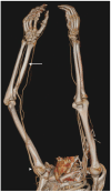
Case: The diagnosis was delayed due to the overlapping cervical radiculopathy. Acute severe pain and nocturnal paresthesia were chief complaints. Ultrasonography, magnetic resonance imaging, and computed tomography angiography revealed that the median nerve was compressed by the occluded median artery. Instead of surgery, conservative therapy was tried. It worked well for six months.
Conclusions: The importance of using modalities for decision making of diagnosis and treatment is emphasized in this report.
Keywords: Carpal tunnel syndrome; Computed tomography angiography; Persistent median artery; Thrombosis.
Copyright © the Korean Society of Anesthesiologists, 2020.
Conflict of interest statement
CONFLICTS OF INTEREST No potential conflict of interest relevant to this article was reported.
Figures


Similar articles
-
Thrombosed persistent median artery causing carpal tunnel syndrome associated with bifurcated median nerve: A case report.Pol J Radiol. 2011 Apr;76(2):46-8. Pol J Radiol. 2011. PMID: 22802832 Free PMC article.
-
Median Nerve Neuropathy Caused by Persistent Median Artery Thrombosis.Plast Reconstr Surg Glob Open. 2023 Apr 10;11(4):e4916. doi: 10.1097/GOX.0000000000004916. eCollection 2023 Apr. Plast Reconstr Surg Glob Open. 2023. PMID: 37359247 Free PMC article.
-
Thrombosis of persistent median artery as a cause of carpal tunnel syndrome : case report.Acta Orthop Belg. 2021 Sep;87(3):529-532. Acta Orthop Belg. 2021. PMID: 34808728
-
[Carpal tunnel syndrome from a thrombosed median artery--four case reports and review of the literature].Handchir Mikrochir Plast Chir. 2009 Jun;41(3):179-82. doi: 10.1055/s-0029-1202839. Epub 2009 Mar 25. Handchir Mikrochir Plast Chir. 2009. PMID: 19322750 Review. German.
-
Radiologic imaging of the carpal tunnel.Eur J Radiol. 1997 Sep;25(2):112-7. doi: 10.1016/s0720-048x(97)00038-7. Eur J Radiol. 1997. PMID: 9283839 Review.
Cited by
-
Carpal tunnel syndrome caused by tophi deposited under the epineurium of the median nerve: A case report.Front Surg. 2023 Jan 6;9:942062. doi: 10.3389/fsurg.2022.942062. eCollection 2022. Front Surg. 2023. PMID: 36684150 Free PMC article.
References
-
- Kopuz C, Baris S, Gulman B. A further morphological study of the persistent median artery in neonatal cadavers. Surg Radiol Anat. 1997;19:403–6. - PubMed
-
- Dahmam A, Matter-Parrat V, Manguila F, Giannikas D, Marin Braun F. Acute carpal tunnel syndrome due to a thrombosed persistent median artery: unusual cause in athletes. J Traumatol Sport. 2015;32:126–8.
-
- Kele H, Verheggen R, Reimers CD. Carpal tunnel syndrome caused by thrombosis of the median artery: the importance of high-resolution ultrasonography for diagnosis. Case report. J Neurosurg. 2002;97:471–3. - PubMed
LinkOut - more resources
Full Text Sources

