Ultrasonography for lumbar neuraxial block
- PMID: 33329842
- PMCID: PMC7724125
- DOI: 10.17085/apm.20065
Ultrasonography for lumbar neuraxial block
Abstract
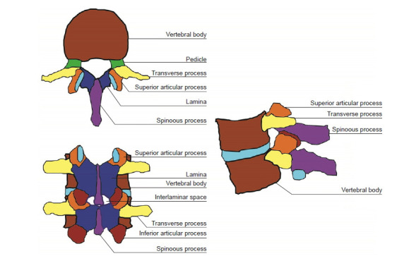
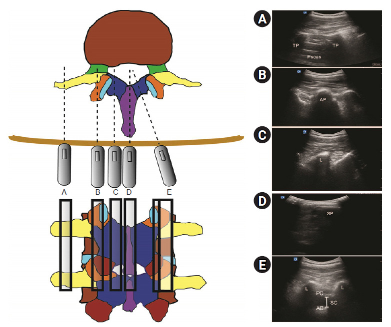
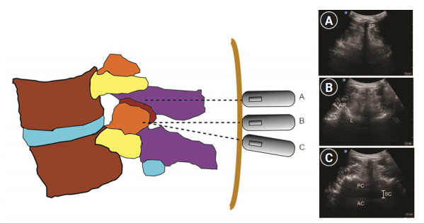
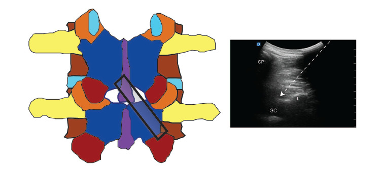
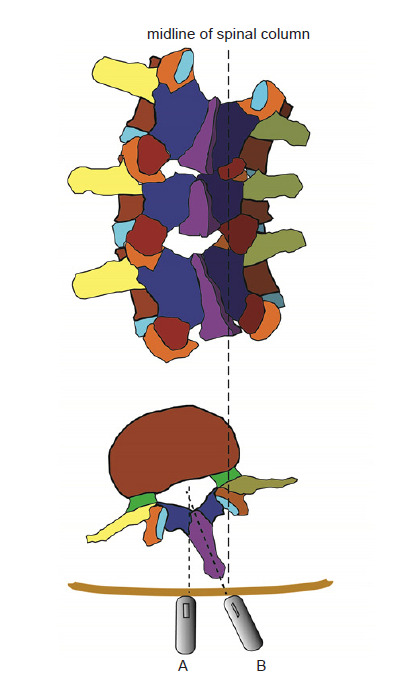
Ultrasonography can be useful to perform a lumbar neuraxial block. It aids in understanding the anatomy of the lumbar spine before the procedure. Pre-procedural ultrasound imaging provides information about the accurate intervertebral level for puncture, optimal needle insertion point, and depth of needle advancement for a successful neuraxial block. The key ultrasonographic views for lumbar neuraxial block include the transverse midline interlaminar and parasagittal oblique views. Ultrasonography can facilitate lumbar neuraxial block in difficult cases, such as the elderly, obese patients, and patients with anatomical abnormality of the lumbar spine. This review elucidates the basics of spinal ultrasonography for lumbar neuraxial block and the current evidence regarding ultrasound-guided neuraxial block in adults.
Keywords: Anesthesia, epidural; Anesthesia, spinal; Lumbar vertebrae; Ultrasonography.
Copyright © the Korean Society of Anesthesiologists, 2020.
Conflict of interest statement
CONFLICTS OF INTEREST No potential conflict of interest relevant to this article was reported.
Figures
References
-
- Chin KJ, Karmakar MK, Peng P. Ultrasonography of the adult thoracic and lumbar spine for central neuraxial blockade. Anesthesiology. 2011;114:1459–85. - PubMed
-
- Pintaric TS, Hadzic A, Strbenc M, Podpecan O, Podbregar M, Cvetko E. Inflammatory response after injection of aqueous gel into subarachnoid space in piglets. Reg Anesth Pain Med. 2013;38:100–5. - PubMed
-
- Killeen T, Kamat A, Walsh D, Parker A, Aliashkevich A. Severe adhesive arachnoiditis resulting in progressive paraplegia following obstetric spinal anaesthesia: a case report and review. Anaesthesia. 2012;67:1386–94. - PubMed
-
- Tran D, Kamani AA, Al-Attas E, Lessoway VA, Massey S, Rohling RN. Single-operator real-time ultrasound-guidance to aim and insert a lumbar epidural needle. Can J Anaesth. 2010;57:313–21. - PubMed
-
- Chin KJ, Chan VW, Ramlogan R, Perlas A. Real-time ultrasound-guided spinal anesthesia in patients with a challenging spinal anatomy: two case reports. Acta Anaesthesiol Scand. 2010;54:252–5. - PubMed
Publication types
LinkOut - more resources
Full Text Sources
Other Literature Sources
Medical