

Neurocysticercosis: An Easy to Miss Diagnosis in Non-Endemic Regions
- PMID: 33329988
- PMCID: PMC7735530
- DOI: 10.7759/cureus.12066
Neurocysticercosis: An Easy to Miss Diagnosis in Non-Endemic Regions
Abstract
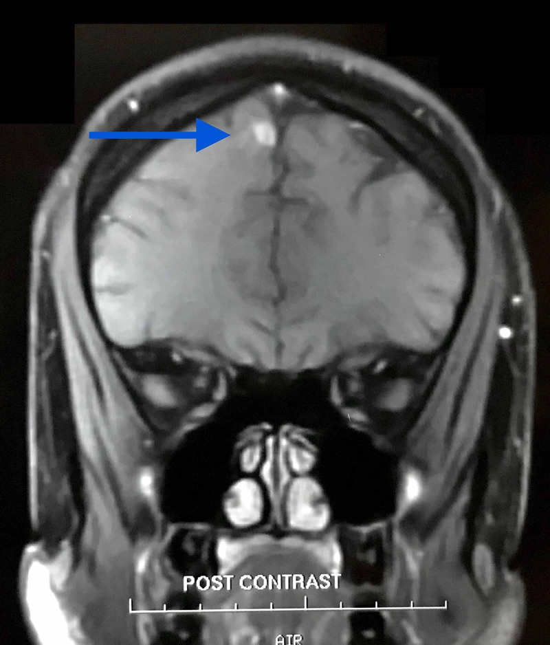
A 29-year-old male presented with swollen gums and stomatitis for the past two months. History revealed that he had moved to the United States from India six years ago and had a first episode of generalized tonic-clonic seizure with confusion and loss of consciousness. Meningioma of the brain was diagnosed, and a Gamma Knife excision of the meningioma was planned. The patient refused to proceed with the surgery and came back to India for a second opinion. Upon repeat MRI scan, the neurosurgeon revised the diagnosis to neurocysticercosis (NCC), and the patient was treated with albendazole, prednisolone, and phenytoin and recovered completely. Hence an unnecessary brain surgery was avoided. The complaint of stomatitis and gingival hypertrophy was due to the side effects of phenytoin. NCC remains a major public health problem in developing countries, and it should be considered as a differential diagnosis in patients from NCC endemic regions.
Keywords: neuro-imaging; neuro-surgery.
Copyright © 2020, Chitkara et al.
Conflict of interest statement
The authors have declared that no competing interests exist.
Figures


References
-
- Epidemiological study of neuro-cysticercosis in northern Togo (West Africa) Dumas M, Grunitzky E, Deniau M, et al. https://pubmed.ncbi.nlm.nih.gov/2488997/ Acta Leiden. 1989;57:191–196. - PubMed
-
- Epidemiology of Taenia solium taeniasis and cysticercosis in two rural Guatemalan communities. Garcia-Noval J, Allan JC, Fletes C, et al. Am J Trop Med Hyg. 1996;55:282–289. - PubMed
-
- Pitfalls in the diagnosis of brain tumours. Omuro AM, Leite CC, Mokhtari K, Delattre JY. Lancet Neurol. 2006;5:937–948. - PubMed
Publication types
LinkOut - more resources
Full Text Sources
Miscellaneous