Evaluation of Muscular Atrophy and Fatty Infiltration Using Time-zero Magnetic Resonance Imaging as Baseline Data, After Rotator Cuff Repair
- PMID: 33330198
- PMCID: PMC7714304
- DOI: 10.5397/cise.2019.22.2.70
Evaluation of Muscular Atrophy and Fatty Infiltration Using Time-zero Magnetic Resonance Imaging as Baseline Data, After Rotator Cuff Repair
Abstract
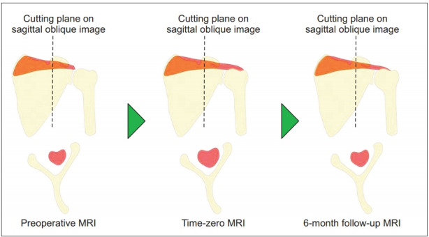
Background: This study evaluated postoperative changes in the supraspinatus from time-zero to 6 months, using magnetic resonance imaging (MRI). We hypothesized that restoration of the musculotendinous unit of the rotator cuff by tendon repair immediately improves the rotator cuff muscle status, and maintains it months after surgery.
Methods: Totally, 76 patients (29 men, 47 women) with rotator cuff tears involving the supraspinatus tendon who underwent arthroscopic rotator cuff repairs were examined. MRI evaluation showed complete repair with intact integrity of the torn tendon at both time-zero and at 6 months follow-up. All patients underwent standardized MRI at our institution preoperatively, at 1 or 2 days postoperative, and at 6 months after surgery. Supraspinatus muscular (SSP) atrophy (Thomazeau grade) and fatty infiltrations (Goutallier stage) were evaluated by MRI. The cross-sectional area of SSP in the fossa was also measured.
Results: As determined by MRI, the cross-sectional area of SSP significantly decreased 11.41% from time-zero (immediate repair) to 6 months post-surgery, whereas the Goutallier stage and Thomazeau grade showed no significant changes (p < 0.01). Furthermore, compared to the preoperative MRI, the postoperative MRI at 6 months showed a no statistically significant increase of 8.03% in the cross-sectional area. In addition, morphological improvements were observed in patients with high grade Goutallier and Thomazeau at time-zero, whereas morphology of patients with low grade factors were almost similar to before surgery.
Conclusions: Our results indicate that cross-sectional area of the initial repair appears to decrease after a few months postoperatively, possibly due to medial retraction or strained muscle.
Keywords: Fatty infiltration; Muscular atrophy; Rotator cuff repair; Time-zero magnetic resonance imaging.
Copyright © 2019 Korean Shoulder and Elbow Society.
Conflict of interest statement
Conflict of interest None.
Figures


References
-
- Goutallier D, Postel JM, Bernageau J, Lavau L, Voisin MC. Fatty infiltration of disrupted rotator cuff muscles. Rev Rhum Engl Ed. 1995;62(6):415–22. - PubMed
-
- Gerber C, Meyer DC, Nuss KM, Farshad M. Anabolic steroids reduce muscle damage caused by rotator cuff tendon release in an experimental study in rabbits. J Bone Joint Surg Am. 2011;93(23):2189–95. doi: 10.2106/JBJS.J.01589. - PubMed
-
- Yoo JC, Ahn JH, Yang JH, Koh KH, Choi SH, Yoon YC. Correlation of arthroscopic repairability of large to massive rotator cuff tears with preoperative magnetic resonance imaging scans. Arthroscopy. 2009;25(6):573–82. doi: 10.1016/j.arthro.2008.12.015. - PubMed
-
- Liem D, Lichtenberg S, Magosch P, Habermeyer P. Magnetic resonance imaging of arthroscopic supraspinatus tendon repair. J Bone Joint Surg Am. 2007;89(8):1770–6. doi: 10.2106/JBJS.F.00749. - PubMed
-
- Rubino LJ, Stills HF, Jr, Sprott DC, Crosby LA. Fatty infiltration of the torn rotator cuff worsens over time in a rabbit model. Arthroscopy. 2007;23(7):717–22. doi: 10.1016/j.arthro.2007.01.023. - PubMed
LinkOut - more resources
Full Text Sources
Research Materials