. 2020 Nov 23:8:566504.
doi: 10.3389/fcell.2020.566504. eCollection 2020.

Histone Demethylase PHF8 Is Required for the Development of the Zebrafish Inner Ear and Posterior Lateral Line

Affiliations

Histone Demethylase PHF8 Is Required for the Development of the Zebrafish Inner Ear and Posterior Lateral Line

Jing He et al. Front Cell Dev Biol. .

Abstract

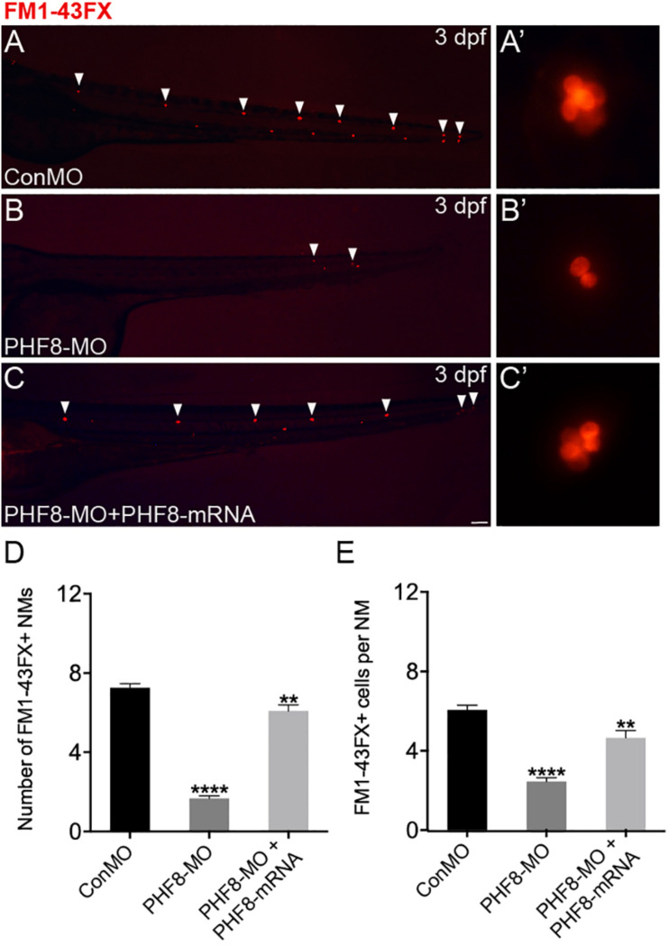
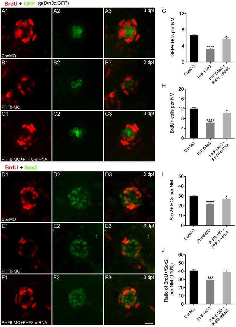
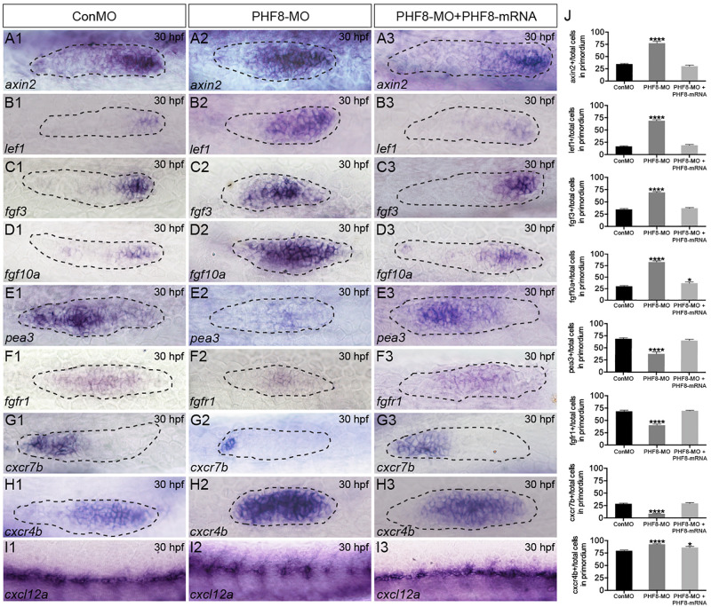
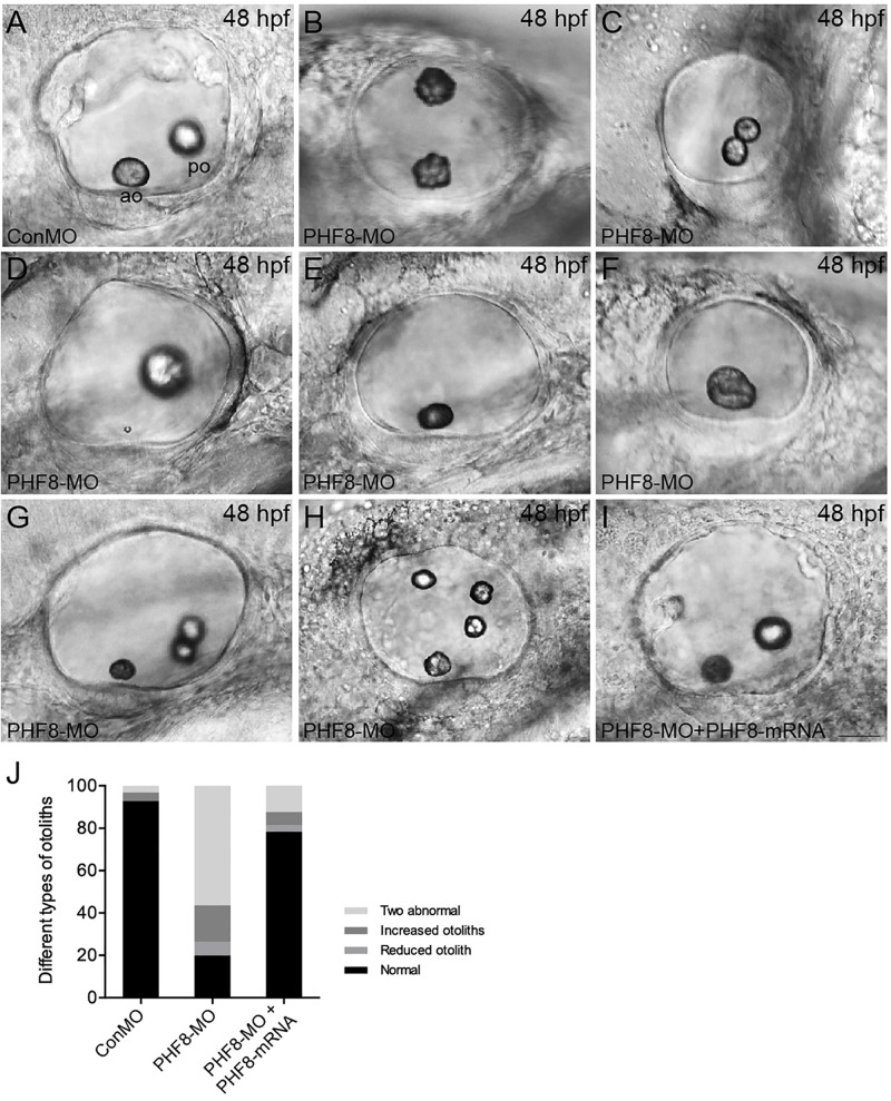
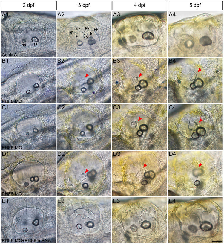
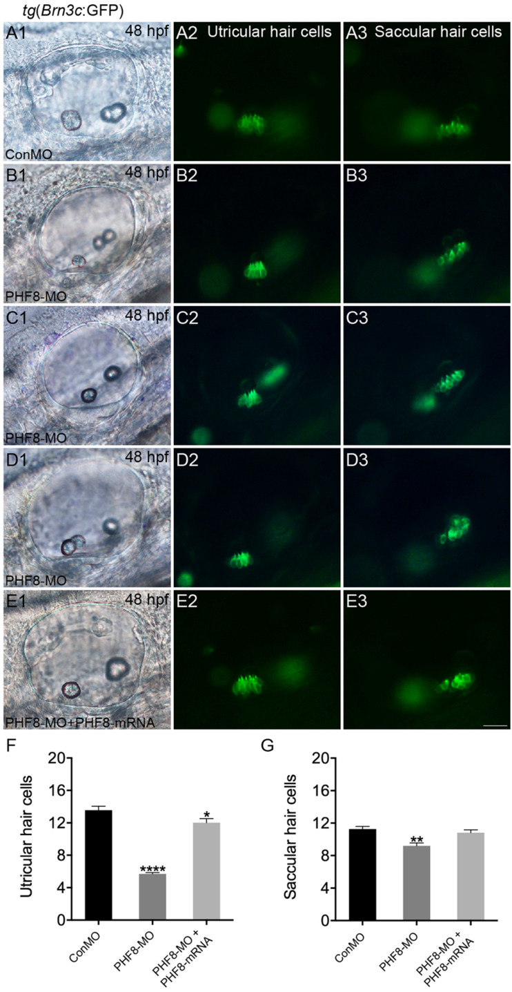
Histone demethylase PHF8 is crucial for multiple developmental processes, and hence, the awareness of its function in developing auditory organs needs to be increased. Using in situ hybridization (ISH) labeling, the mRNA expression of PHF8 in the zebrafish lateral line system and otic vesicle was monitored. The knockdown of PHF8 by morpholino significantly disrupted the development of the posterior lateral line system, which impacted cell migration and decreased the number of lateral line neuromasts. The knockdown of PHF8 also resulted in severe malformation of the semicircular canal and otoliths in terms of size, quantity, and position during the inner ear development. The loss of function of PHF8 also induced a defective differentiation in sensory hair cells in both lateral line neuromasts and the inner ear. ISH analysis of embryos that lacked PHF8 showed alterations in the expression of many target genes of several signaling pathways concerning cell migration and deposition, including the Wnt and FGF pathways. In summary, the current findings established PHF8 as a novel epigenetic element in developing auditory organs, rendering it a potential candidate for hearing loss therapy.

Keywords: PHF8; inner ear; organogenesis; posterior lateral line; zebrafish.


Figures

FIGURE 1
FIGURE 1
Transcription patterns of Phf8 from 2-cell stage to 48 hpf. (A,B) Phf8 expression was detected in both blastomeres at the 2-cell stage and in whole blastoderm at 16-cell stage. (C–F) Expression of Phf8 in PLL system and otic vesicle at 32 and 48 hpf. (C1,C2; PLLp, 32 hpf), neuromasts (E1,E2; NM, 48 hpf), and otic vesicle (D1,D2,F1,F2; OV, 32 and 48 hpf). Black arrows indicate the deposited neuromast, the white asterisks show protrusions of the semicircular canal, and the white square bracket shows the cristae. PLL primordium, PLLp; Neuromast, NM; otic vesicle, OV. Arrows.
FIGURE 2
FIGURE 2
Abnormal PLL neuromast deposition in the PHF8 deficient embryos. (A–F) Optical appearance of fluorescence of 48 hpf larvae expressing Et(gata2:EGFP)mp189b. (A) The standard control MO (ConMO)-injected embryo illustrates a completed PLL mode. (B–C) PHF8-deficient embryos demonstrate a decrease in deposited neuromasts. (D) The embryo co-injected with PHF8-MO and p53-MO. (E) The embryo is injected with PHF8-mRNA. (F) The embryo co-injected with PHF8-MO and PHF8-mRNA. White arrowheads implicate the positions of the neuromasts on one side, Red arrowhead implicates the location of PLL primordium. (G,H) The quantity of PLL neuromasts (G) and the position of the first neuromast (NM1; H) were measured. Control embryos (ConMO; n = 112 embryos), PHF8 morphants (PHF8-MO; n = 319 embryos), PHF8-MO co-injected with P53-MO embryos (PHF8-MO + P53-MO; n = 115 embryos), PHF8 embryos injected with mRNA (PHF8-mRNA; n = 76 embryos), and PHF8-MO co-injected with PHF8-mRNA embryos (PHF8-MO + PHF8-mRNA; n = 80 embryos). **p < 0.01,****p < 0.0001 vs. the ConMO group. Data are presented as means ± SEM. All pictures showed lateral view, anterior left. Scale bars, 100 μm. Neuromast, NM.
FIGURE 3
FIGURE 3
Analysis of neuromast hair cells in PHF8 morphant. (A–C) Embryos from different groups were stained with FM1-43FX at 3 dpf to detect functional neuromasts. (A’–C’) The enlarged FM1-43FX-positive neuromasts in controls (A’), PHF8 morphants (B’), and PHF8-MO co-injected with PHF8-mRNA embryos (C’) at 3 dpf. (D) The average number of FM1-43FX-positive neuromasts in controls (ConMO; n = 12 embryos), PHF8 morphants (PHF8-MO; n = 12 embryos), and PHF8-MO co-injected with PHF8-mRNA embryos (PHF8-MO + PHF8-mRNA; n = 12 embryos) at 3 dpf. (E) The average number of FM1-43FX-positive cells per neuromast (NM) in controls (ConMO; n = 20 neuromasts), PHF8 morphants (PHF8-MO; n = 20 neuromasts), and PHF8-MO co-injected with PHF8-mRNA embryos (PHF8-MO + PHF8-mRNA; n = 20 neuromasts) at 3 dpf. White arrowheads implicate the locations of the neuromasts on one side. **p < 0.01, ****p < 0.0001 vs. the ConMO group. Data are presented as means ± SEM. All pictures showed lateral view, anterior left. Scale bar, 100 μm. Neuromast, NM.
FIGURE 4
FIGURE 4
Analysis of neuromast proliferation in PHF8 morphant. (A–C) Representative images of GFP (green) and BrdU (red) staining of neuromasts from control (ConMO; A), PHF8 morphants (PHF8-MO; B), and PHF8-MO co-injected with PHF8-mRNA embryo (PHF8-MO + PHF8-mRNA; C) at 3 dpf. Hair cells were assessed with GFP visualization in transgenic line tg(Brn3c:GFP). (D–F) Representative images of supporting cell marker Sox2 (green) and BrdU (red) staining of neuromasts from control (ConMO; D), PHF8 morphants (PHF8-MO; E), and PHF8-MO co-injected with PHF8-mRNA embryo (PHF8-MO + PHF8-mRNA; F) at 3 dpf. (G–J) The average number of GFP-positive hair cells (G), BrdU-positive cells (H), Sox2-positive supporting cells (I), and the ratio of BrdU + /Sox2 + cells per neuromast (NM) in control (ConMO), PHF8 morphants (PHF8-MO), and PHF8-MO co-injected with PHF8-mRNA embryo (PHF8-MO + PHF8-mRNA) at 3 dpf. n = 24 neuromasts per group, *p < 0.05, ***p < 0.001, ****p < 0.0001 vs. the ConMO group. Statistics are presented as means ± SEM. Scale bar, 10 μm. Neuromast, NM.
FIGURE 5
FIGURE 5
Analysis of expression of genes of Wnt and FGF signaling pathways, as well as cxcl12/cxc7b/cxcr4b. (A–I) WISH was utilized to examine the expression of axin2 (A), lef1 (B), fgf3 (C), fgf10a (D), pea3 (E), fgfr1 (F), cxcr7b (G), cxcr4b (H), and cxcl12 (I) at 30 hpf. (J) The ratio of axin2, lef1, fgf3, fgf10a, pea3, fgfr1, cxcr7b, and cxcr4b-positive cells to total cells in the primordia of controls (ConMO; n = 10 embryos for axin2, 10 embryos for lef1, 12 embryos for fgf3, 12 embryos for fgf10a, 11 embryos for pea3, 9 embryos for fgfr1, 10 embryos for cxcr7b, 12 embryos for cxcr4b), PHF8 morphants (PHF8-MO; n = 10 embryos for axin2, 10 embryos for lef1, 14 embryos for fgf3, 17 embryos for fgf10a, 9 embryos for pea3, 9 embryos for fgfr1, 15 embryos for cxcr7b, 17 embryos for cxcr4b), and PHF8-MO co-injected with PHF8-mRNA embryo (PHF8-MO + PHF8-mRNA; n = 10 embryos for axin2, 10 embryos for lef1, 10 embryos for fgf3, 9 embryos for fgf10a, 9 embryos for pea3, 10 embryos for fgfr1, 12 embryos for cxcr7b, 12 embryos for cxcr4b). Data are expressed as mean ± SEM. *p < 0.05, ****p < 0.0001. All optical appearances elucidate lateral visions; anterior is on the left. The primordium is delineated with a dashed line.
FIGURE 6
FIGURE 6
Analysis of otic vesicle formation in PHF8 morphant. (A–I) The overall morphology of otic vesicles in control embryos (ConMO; A), PHF8 morphants (PHF8-MO; B–H), and PHF8-MO co-injected with PHF8-mRNA embryos (PHF8-MO + PHF8-mRNA; I) at 48 hpf. According to the otolith phenotypes, zebrafish PHF8 morphants were divided into types as follows: the 2 abnormal otolith groups (B, misplaced; C, fused; D, tiny); the 1 otolith group (E,F); and the multiple otolith group (G,H). (J) Statistical results of different kinds of malformed otoliths (n = 125 for ConMO embryos, n = 126 for PHF8 morphants, and n = 75 for PHF8-MO + PHF8-mRNA embryos). The positions of the anterior otolith (ao) and posterior otolith (po) are implied. Optical appearances elucidate lateral visions; anterior is on the left. Scale bar, 25 μm.
FIGURE 7
FIGURE 7
Analysis of semicircular canal formation in PHF8 morphant. (A–E) The overall morphology of semicircular canal in control embryos (ConMO; A), PHF8 morphants (PHF8-MO; B–D), and PHF8-MO co-injected with PHF8-mRNA embryos (PHF8-MO + PHF8-mRNA; E) at 2 dpf (A1–E1), 3 dpf (A2–E2), 4 dpf (A3–E3), and 5 dpf (A4–E4). Black arrowheads indicate the junction of the anterior protrusion (ap) and the anterior bulge (ab) and the junction of the posterior bulge (pb) and posterior protrusion (pp). Asterisks point out the junction between the ventral bulge (vb) and ventral protrusion (vp). Red arrowheads mark disrupted projections. The locations of the anterior otolith (ao) and posterior otolith (po) are implied. All pictures are lateral views with the anterior to the left and the dorsal side up. Scale bar, 50 μm.
FIGURE 8
FIGURE 8
The requirement of PHF8 for hair cells in the inner ear. (A–D) Representative images of GFP (green) in the utricle and saccule of control embryo (ConMO; A1–A3), PHF8 morphant (PHF8-MO; B1–B3,C1–C3,D1–D3), and PHF8-MO co-injected with PHF8-mRNA embryos (PHF8-MO + PHF8-mRNA; E1–E3) at 48 hpf. Hair cells were assessed with GFP visualization in transgenic line tg(Brn3c:GFP). (F–G) Quantification of the numbers of hair cells in the utricle and saccule of the inner ear for each experimental condition at 48 hpf. *p < 0.05, **p < 0.01, ****p < 0.0001 vs. the ConMO group. Data are presented as means ± SEM (n = 16 embryos, per each). Optical appearances elucidate lateral visions; anterior is on the left. Scale bar, 25 μm.

References

    1. Abidi F. E., Miano M. G., Murray J. C., Schwartz C. E. (2007). A novel mutation in the PHF8 gene is associated with X-linked mental retardation with cleft lip/cleft palate. Clin. Genet. 72 19–22. 10.1111/j.1399-0004.2007.00817.x - DOI - PMC - PubMed
    1. Aman A., Piotrowski T. (2008). Wnt/beta-catenin and Fgf signaling control collective cell migration by restricting chemokine receptor expression. Dev. Cell 15 749–761. 10.1016/j.devcel.2008.10.002 - DOI - PubMed
    1. Bever M. M., Fekete D. M. (2002). Atlas of the developing inner ear in zebrafish. Dev. Dyn. 223 536–543. 10.1002/dvdy.10062 - DOI - PubMed
    1. Dambly-Chaudiere C., Sapede D., Soubiran F., Decorde K., Gompel N., Ghysen A. (2003). The lateral line of zebrafish: a model system for the analysis of morphogenesis and neural development in vertebrates. Biol. Cell 95 579–587. 10.1016/j.biolcel.2003.10.005 - DOI - PubMed
    1. David N. B., Sapede D., Saint-Etienne L., Thisse C., Thisse B., Dambly-Chaudiere C., et al. (2002). Molecular basis of cell migration in the fish lateral line: role of the chemokine receptor CXCR4 and of its ligand, SDF1. Proc. Natl. Acad. Sci. U.S.A. 99 16297–16302. 10.1073/pnas.252339399 - DOI - PMC - PubMed

LinkOut - more resources