Review
. 2020 Oct 29:7:544302.
doi: 10.3389/fcvm.2020.544302. eCollection 2020.

Arterial Stiffness and Hypertension in the Elderly

Affiliations
Review

Arterial Stiffness and Hypertension in the Elderly

Stéphane Laurent et al. Front Cardiovasc Med. .

Abstract

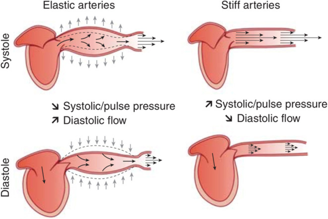
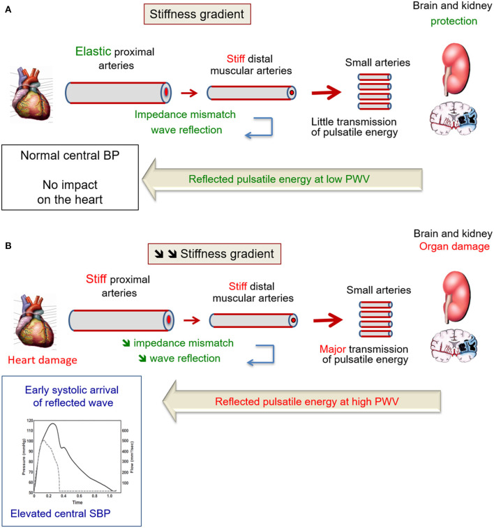
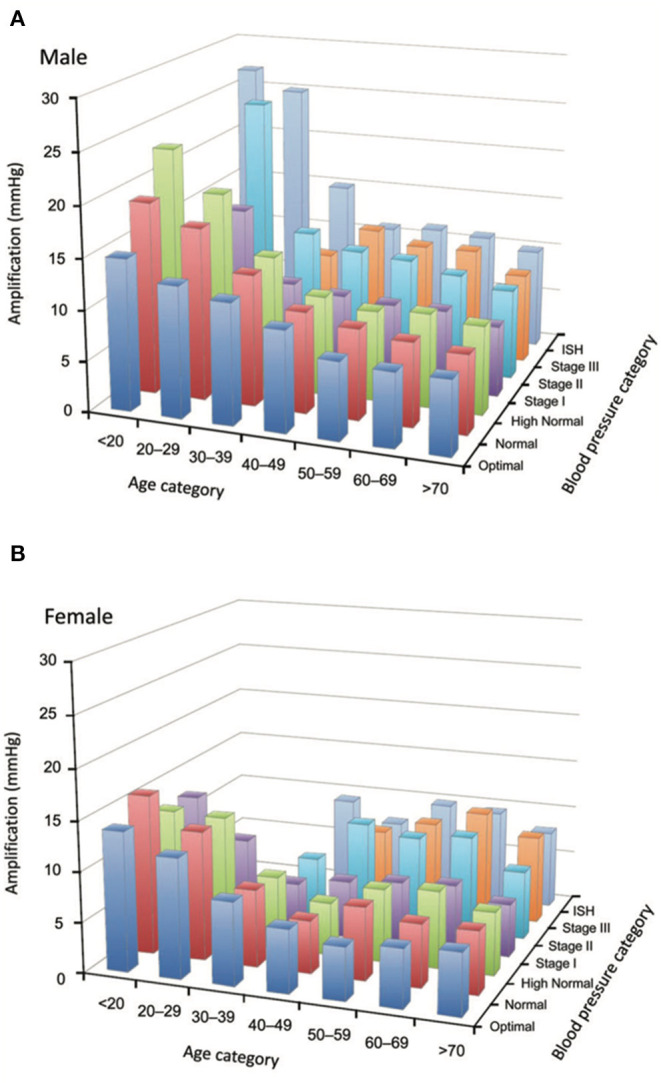
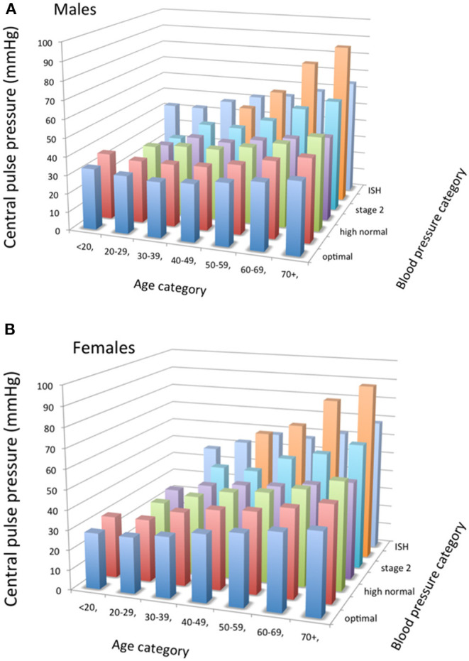
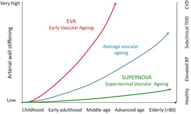
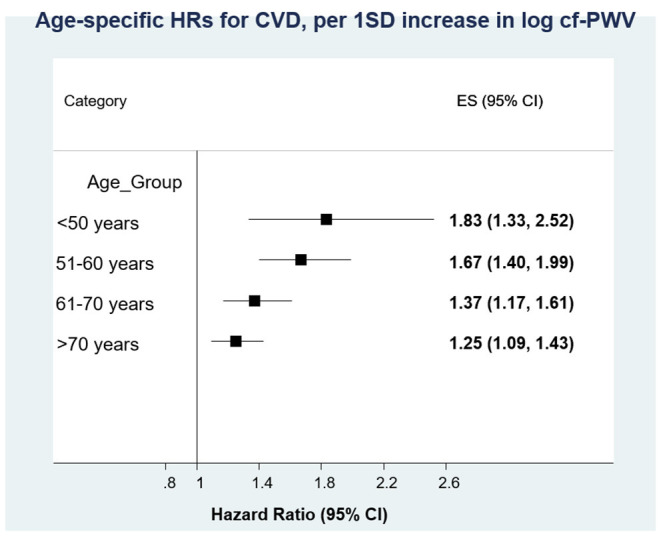
Hypertension prevalence increases with age. Age and high blood pressure are the two main determinants of arterial stiffness. In elderly hypertensives, large arteries stiffen and systolic and pulse pressures increase, due to wave reflections. A major reason for measuring arterial stiffness in clinical practice in elderly hypertensive patients comes from the repeated demonstration that arterial stiffness and wave reflections have a predictive value for CV events. A large body of evidence has been published during the last two decades, concerning the epidemiology, pathophysiology, and pharmacology of large arteries in hypertension in various settings of age. Particularly, two expert consensus documents have reviewed the methodological agreements for measuring arterial stiffness. The concepts of Early Vascular Aging (EVA) and Supernormal Vascular Aging (SUPERNOVA) help to better understand on which determinants of arterial stiffness it is possible to act, in order to limit target organ damage and cardiovascular complications. This review will address the issues of the cellular and molecular mechanisms of arterial stiffening in elderly hypertensives, the consequences of arterial stiffening on central systolic and pulse (systolic minus diastolic, PP) pressures and target organs, the methodology for measuring arterial stiffness, central pulse pressure and wave reflection, the epidemiological determinants of arterial stiffening in elderly hypertensives, the pharmacology of arterial destiffening, and how the concepts of EVA and SUPERNOVA apply to the detection of organ damage and prevention of CV complications.

Keywords: age; arterial stiffness; cardiovascular events; central blood pressure; elderly; large artery; organ damage.


Figures

Figure 1
Figure 1
Schematic representation of the role of arterial compliance (i.e., the inverse of arterial stiffness) in dampening blood pressure pulsatility and assuring adapted blood flow through the peripheral circulation. From Briet et al. (20) with permission.
Figure 2
Figure 2
(A) Healthy aging is a means to retard brain, kidney, and heart damage. Indeed, in healthy subjects, an impedance mismatch occurs in response to the stiffness gradient between proximal elastic arteries and distal muscular arteries. This phenomenon generates pressure wave reflection, limiting the transmission of pressure pulsatility to target organs. The largest part of the reflected pulsatile energy that propagates backward, toward the heart, travels indeed at low velocity along elastic arteries, thus do not superimpose on incident pressure wave. Thus, central BP remains normal. From Laurent and Cunha (23) with permission. (B) Wave reflections in the elderly. When the aorta stiffens with aging, it loses its ability to dampen the pulsatility of ventricular ejection. Small arteries of target organ are damaged by the hyperpulsatility. Because the stiffness of distal muscular arteries does not change with age, there is a reduction of the stiffness gradient between proximal elastic arteries and distal muscular arteries. Thus, pressure pulsatility is transmitted to a larger extent toward small arteries of target organs. The largest part of the reflected pulsatile energy that propagates backward, toward the heart, travels at high velocity along stiff arteries. TRhus, it superimposes on incident pressure wave and increase central SBP. From Laurent and Cunha (23) with permission.
Figure 3
Figure 3
(A,B) Loss of amplification phenomenon in the elderly. Tridimensional bar-graphs representing amplification according to sex (6a, males; 6b, females), age categories, and blood pressure categories. The value represented here is the median of the group. Some categories are not represented because there were <50 observations. From Herbert et al. (32) with permission.
Figure 4
Figure 4
(A,B) Tridimensional bar-graphs representing central pulse pressure (peripheral minus central systolic blood pressures) according to sex (7a, males; 7b, females), age categories, and blood pressure categories. The value represented here is the median of the group. Some categories are not represented because there were <50 observations. From Herbert et al. (32), with permission.
Figure 5
Figure 5
This figure illustrates, in a life-course approach of hypertension, our hypothesis that progressive arterial wall stiffening with aging parallels incident hypertension, and then subclinical target organ damage, and then CV complications. EVA subjects reach each of these steps earlier than the average population, whereas SUPERNOVA subjects remain protected for a long period of time.
Figure 6
Figure 6
Measurement of carotid-femoral pulse wave velocity with the foot-to-foot method. From Laurent et al. (1), with permission. The waveforms are usually obtained transcutaneously at the right common carotid artery and the right femoral artery. The time delay (Δt, or transit time) is measured between the feet of the two waveforms. The distance (ΔL) covered by the waves is usually assimilated to the surface distance between the two recording sites, i.e., the common carotid artery and the common femoral artery. PWV is calculated as PWV = 0.8 × ΔL (meters)/Δt (seconds).
Figure 7
Figure 7
Individual participant systematic review of the predictive value of arterial stiffness (carotid-femoral pulse wave velocity) for cardiovascular disease. Associations stratified according to sex, diabetes, and hypertension were similar but decreased with age, with a significant interaction. From Ben Shlomo et al. (4), with permission.

References

    1. Laurent S, Cockcroft J, Van Bortel L, Boutouyrie P, Giannattasio C, Hayoz D, et al. . Expert consensus document on arterial stiffness: methodological aspects and clinical applications. Eur Heart J. (2006) 27:2588–605. 10.1093/eurheartj/ehl254 - DOI - PubMed
    1. Vlachopoulos C, Aznaouridis K, Stefanadis C. Prediction of cardiovascular events and all-cause mortality with arterial stiffness: a systematic review and meta-analysis. J Am Coll Cardiol. (2010) 55:1318–27. 10.1016/j.jacc.2009.10.061 - DOI - PubMed
    1. Vlachopoulos C, Aznaouridis K, O'Rourke MF, Safar ME, Baou K, Stefanadis C. Prediction of cardiovascular events and all-cause mortality with central haemodynamics: a systematic review and meta-analysis. Eur Heart J. (2010) 31:1865–71. 10.1093/eurheartj/ehq024 - DOI - PubMed
    1. Ben-Shlomo Y, Spears M, Boustred C, May M, Anderson SG, Benjamin EJ, et al. . Aortic pulse wave velocity improves cardiovascular event prediction: an individual participant meta-analysis of prospective observational data from 17,635 subjects. J Am Coll Cardiol. (2014) 63:636–46. 10.1016/j.jacc.2013.09.063 - DOI - PMC - PubMed
    1. van Bortel LM, Laurent S, Boutouyrie P, Chowienczyk P, Cruickshank JK, de Backer T, et al. . Expert consensus document on the measurement of aortic stiffness in daily practice using carotid-femoral pulse wave velocity. J Hypertens. (2012) 30:445–8. 10.1097/HJH.0b013e32834fa8b0 - DOI - PubMed