[Preprint]. 2021 Nov 11:2020.12.08.415505.
doi: 10.1101/2020.12.08.415505.

Thiol drugs decrease SARS-CoV-2 lung injury in vivo and disrupt SARS-CoV-2 spike complex binding to ACE2 in vitro

Affiliations

Thiol drugs decrease SARS-CoV-2 lung injury in vivo and disrupt SARS-CoV-2 spike complex binding to ACE2 in vitro

Kritika Khanna et al. bioRxiv. .

Abstract

Neutrophil-induced oxidative stress is a mechanism of lung injury in COVID-19, and drugs with a functional thiol group ("thiol drugs"), especially cysteamine, have anti-oxidant and anti-inflammatory properties that could limit this injury. Thiol drugs may also alter the redox status of the cysteine-rich SARS-CoV-2 spike glycoprotein (SARS-2-S) and thereby disrupt ACE2 binding. Using ACE2 binding assay, reporter virus pseudotyped with SARS-CoV-2 spikes (ancestral and variants) and authentic SARS-CoV-2 (Wuhan-1), we find that multiple thiol drugs inhibit SARS-2-S binding to ACE2 and virus entry into cells. Pseudoviruses carrying variant spikes were less efficiently inhibited as compared to pseudotypes bearing an ancestral spike, but the most potent drugs still inhibited the Delta variant in the low millimolar range. IC50 values followed the order of their cystine cleavage rates and lower thiol pKa values. In hamsters infected with SARS-CoV-2, intraperitoneal (IP) cysteamine decreased neutrophilic inflammation and alveolar hemorrhage in the lungs but did not decrease viral infection, most likely because IP delivery could not achieve millimolar concentrations in the airways. These data show that thiol drugs inhibit SARS-CoV-2 infection in vitro and reduce SARS-CoV-2-related lung injury in vivo and provide strong rationale for trials of systemically delivered thiol drugs as COVID-19 treatments. We propose that antiviral effects of thiol drugs in vivo will require delivery directly to the airways to ensure millimolar drug concentrations and that thiol drugs with lower thiol pKa values are most likely to be effective.


Conflict of interest statement

Competing interests: John Fahy, Stefan Oscarson, Irina Gitlin and Wilfred Raymond are inventors on patent applications related to use of thiol-based drugs as treatments for COVID19. The other authors have no competing interests.

Figures
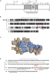
Fig. 1.
Fig. 1.. Cystine mapping and conservation of cystines in RBD of SARS-1-S and SARS-2-S.
(A) Cystine map for SARS-2-S domain S1 (UniProt: P0DTC2). Ten cystine linkages are denoted by dashed lines with amino acid residue number above. The dark gray region is the RBD, and the lighter gray box highlights the ACE2 binding motif, a cluster of amino acids that make contact with ACE2. (B) Amino acid alignment of SARS-2-S RBD domain (aa 319–541, PDB: 6M0J) and SARS-1-S RBD domain (aa 306–517, PDB: 3SCI). Residues that are shared are highlighted by black boxes and residues that represent a similar amino acid class replacement are bound by gray boxes. The solid lines link cystine-forming cysteines. The solid red line and red numbers highlight the conserved critical cystine bridge in the RBDs for both viruses. Asterisks denote amino acids that are within 4 angstroms of ACE2 in their respective solved structures. (C) A surface rendering of SARS-2-RBD (PDB: 6M0J) oriented with the ACE2 binding region (blue) facing forward. (D) Amino acid sequence of SARS-2-S RBD highlighting the RBD mutations identified in the circulating SARS-CoV-2 variants. Amino acids are noted with single letter code and sequence number. The conserved RBD cystine (C480 -C488) is highlighted in red.
Fig. 2.
Fig. 2.. Binding of SARS-CoV-2 RBD to ACE2 is inhibited by thiol-based drugs.
(A) Schematic representation of the SARS-CoV-2 RBD to ACE2 binding assay. (B) Percent of binding of RBDWuhan−1 to ACE2 in the presence of the drugs (n = 4–6). (C) Area under the curve (AUC) analysis of (B). (D) Binding of RBDWuhan−1 to ACE2 at one and two hours post drug exposure and washout (n = 4 – 5). Reference AUC was calculated from RBD to ACE2 binding with no drug control; dashed line represents 50% of reference AUC. Statistical significance for (C) was analyzed by one-way ANOVA followed by Dunnett’s post-hoc analysis. Significance indicates differences from reference AUC. **p ≤ 0.01, ****, p ≤ 0.0001.
Fig. 3.
Fig. 3.. Thiol-based drugs inhibit SARS-CoV-2 in vitro.
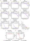
(A to H) Pseudovirus (PV) entry efficiency after pseudovirus exposure to (A) carbocysteine (sulfide drug, negative control), (B) amifostine (parent drug of WR-1065, negative control), thiol-based drugs (C) NAC, (D) Tiopronin, (E) Mesna, (F) Bucillamine, (G) WR-1065, and (H) Cysteamine prior to transduction into 293T-ACE2-TMPRSS2 cells (n = 3–4). The effects of drugs on cell viability were quantified using Cell Titer Glo 2.0 with lower drug dose exposures, reflecting 66-fold dilution of drugs when pseudovirus/drug mixture was incubated with cells (n=3). (I) IC50 values of the different thiol drugs in the PV assay. (J-K) Cytopathic effects (CPE) quantified by visual inspection when authentic SARS-CoV-2 is exposed to (J) carbocysteine (negative control) and (K) cysteamine (thiol drug) prior to infection in Vero E6-TMPRSS2 cells (n = 3). The effects of drugs on cell viability were quantified with exposure of cells to lower drug doses, reflecting the 24-fold dilution of drugs when virus/drug mixture was incubated with cells (n=3). The X-axes are scaled to log10− the lower X-axis refers to the concentration of drugs on the PV/ live virus and the upper X-axis refers to concentration of drugs on the cells post dilution. Percentage changes are with respect to no drug control which is set as 100%. IC50 of the drugs was determined using the non-linear regression fitting with a variable slope. Data are mean ± SD.
Fig. 4.
Fig. 4.. Thiol drugs reduce the infection of 293T-ACE2-TMPRSS2 cells by SARS-CoV-2 variant pseudoviruses
(A to F) Pseudovirus (PV) entry efficiency when the B.1.617.1 and B.1.617.2 pseudoviruses were exposed to (A) carbocysteine (sulfide drug, negative control), thiol drugs (B) Mesna, (C) Bucillamine, (D) TDG, (E) Cysteamine and (F) WR-1065 prior to transduction into 293T-ACE2-TMPRSS2 cells (n = 3–4). (G) IC50 values of the thiol drugs in the B.1.617.1 and B.1.617.2 PV transduction assay and their respective thiol pKa values. (H, I) Pseudovirus (PV) entry efficiency when the SARS-CoV-2 variant pseudoviruses were exposed to (H) carbocysteine (sulfide drug, negative control) and thiol drug (I) cysteamine prior to transduction into 293T-ACE2-TMPRSS2 cells (n = 3–4). (J) IC50 values of cysteamine in the SARS-CoV-2 variant PV transduction assays. NA = Not achieved.
Fig. 5.
Fig. 5.. Thiol pKa influences the cystine cleaving rates of the thiol drugs and their efficacy in B.1.617.1 PV transduction assay.
(A to C) PV entry efficiency when B.1.617.1 pseudoviruses were treated with three cysteine derivates (A) N-acetyl cysteine, (B) L-cysteine and (C) L-cysteine methyl ester (CME) having variable thiol pKa (n=3–6). Data are mean ± SD. (D) Schematic representation of the BODIPY cystine assay. (E) Rate of BODIPY FL-cystine cleavage when exposed to thiol drugs or controls (carbocysteine, amifostine) (n=3). Dotted lines indicate SEM. (F) Maximum slope (Max V) for the reactivity of thiol drugs with the BODIPY FL cystine reagent [based on data in (E)]. Numbers on graph (E) correspond with numbers designated to thiol compounds in (F). Data in (F) are mean ± SEM.
Figure 6:
Figure 6:. Effects of cysteamine on a Syrian hamster model of SARS-CoV-2 infection.
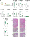
(A) Study design for assessing the effect of cysteamine in Syrian hamster model of COVID-19. Cysteamine (100mg/kg) was administered twice daily via intraperitoneal injection for 5 days, with the first dose given 2 hours prior to the virus inoculation on Day 0. (B) Viral RNA levels in the lungs of animals treated with cysteamine relative to the vehicle control group. (C) Lung weights, normalized to the terminal body weights, of the animals. Total protein (D) and total cell counts (E) in the BAL fluid of hamsters treated with cysteamine with respect to the vehicle controls. (F-I) Differential leukocyte counts in the BAL fluid of animals, showing (F) neutrophil, (G) macrophage, (H) lymphocyte and (I) eosinophil counts in treated and vehicle control groups. (J, K) Histopathology scores for mixed cell (macrophages and neutrophils) inflammation in the peribronchovascular and the centriacinar regions of the lung (J) and alveolar hemorrhage (K) in the lungs of animals treated with cysteamine relative to vehicle control group. (L) Representative images of the lung sections of the animals highlighting the extent of alveolar hemorrhage in the animals from the two groups. Scale bar for 4X images equal 200μm; scale bar for 10X images equal 500μm. Control – intraperitoneal vehicle control group; CYS - cysteamine. Each group had N=10 animals. 2 BAL samples from the cysteamine group could not be analyzed due to a technical error. Data are mean ± SEM. Statistical significance was analyzed by two tailed, unpaired t-test between cysteamine treated (CYS) and respective control group (control).

References

    1. Wiersinga W. J., Rhodes A., Cheng A. C., Peacock S. J., Prescott H. C., Pathophysiology, Transmission, Diagnosis, and Treatment of Coronavirus Disease 2019 (COVID-19): A Review JAMA - J. Am. Med. Assoc. (2020), doi:10.1001/jama.2020.12839. - DOI - PubMed
    1. Zhu N., Zhang D., Wang W., Li X., Yang B., Song J., Zhao X., Huang B., Shi W., Lu R., Niu P., Zhan F., Ma X., Wang D., Xu W., Wu G., Gao G. F., Tan W., A novel coronavirus from patients with pneumonia in China, 2019, N. Engl. J. Med. 382, 727–733 (2020). - PMC - PubMed
    1. Laforge M., Elbim C., Frère C., Hémadi M., Massaad C., Nuss P., Benoliel J.-J., Becker C., Tissue damage from neutrophil-induced oxidative stress in COVID-19, Nat. Rev. Immunol. 2020 209 20, 515–516 (2020). - PMC - PubMed
    1. Horby P., Lim W. S., Emberson J. R., Mafham M., Bell J. L., Linsell L., Staplin N., Brightling C., Ustianowski A., Elmahi E., Prudon B., Green C., Felton T., Chadwick D., Rege K., Fegan C., Chappell L. C., Faust S. N., Jaki T., Jeffery K., Montgomery A., Rowan K., Juszczak E., Baillie J. K., Haynes R., Landray M. J., Dexamethasone in Hospitalized Patients with Covid-19, N. Engl. J. Med. 384, 693–704 (2021). - PMC - PubMed
    1. R. C. Group, Tocilizumab in patients admitted to hospital with COVID-19 (RECOVERY): a randomised, controlled, open-label, platform trial, Lancet (London, England) 397, 1637–1645 (2021). - PMC - PubMed

Publication types