. 2021 Jan 11;49(1):306-321.
doi: 10.1093/nar/gkaa1188.

An atypical BRCT-BRCT interaction with the XRCC1 scaffold protein compacts human DNA Ligase IIIα within a flexible DNA repair complex

Affiliations

An atypical BRCT-BRCT interaction with the XRCC1 scaffold protein compacts human DNA Ligase IIIα within a flexible DNA repair complex

Michal Hammel et al. Nucleic Acids Res. .

Abstract

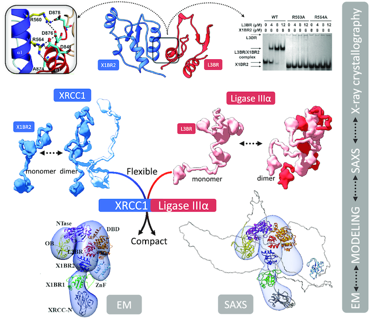
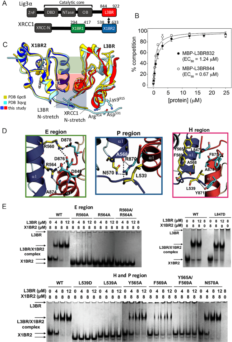
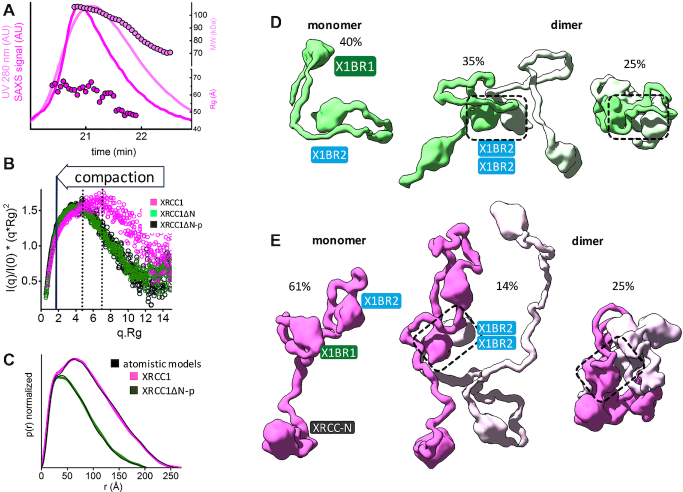
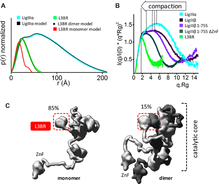
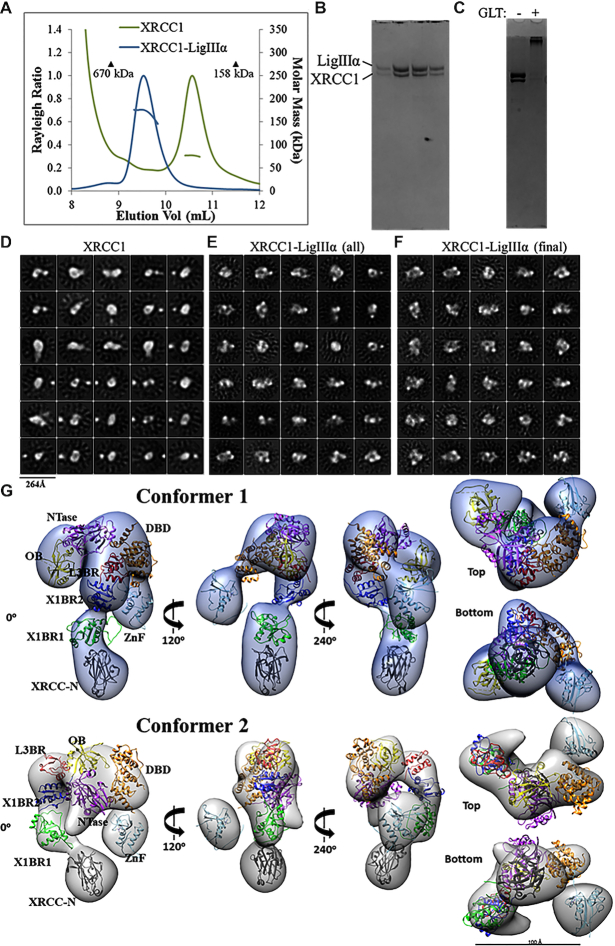
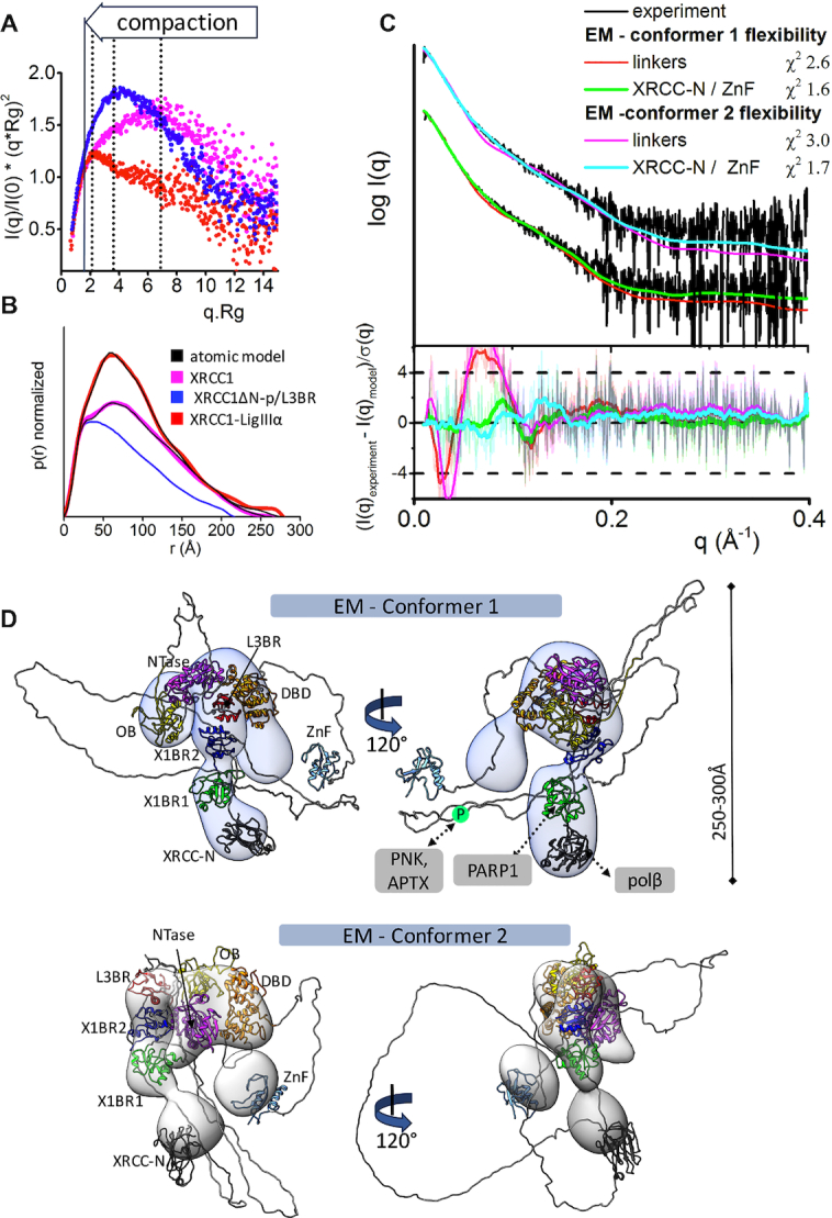
The XRCC1-DNA ligase IIIα complex (XL) is critical for DNA single-strand break repair, a key target for PARP inhibitors in cancer cells deficient in homologous recombination. Here, we combined biophysical approaches to gain insights into the shape and conformational flexibility of the XL as well as XRCC1 and DNA ligase IIIα (LigIIIα) alone. Structurally-guided mutational analyses based on the crystal structure of the human BRCT-BRCT heterodimer identified the network of salt bridges that together with the N-terminal extension of the XRCC1 C-terminal BRCT domain constitute the XL molecular interface. Coupling size exclusion chromatography with small angle X-ray scattering and multiangle light scattering (SEC-SAXS-MALS), we determined that the XL is more compact than either XRCC1 or LigIIIα, both of which form transient homodimers and are highly disordered. The reduced disorder and flexibility allowed us to build models of XL particles visualized by negative stain electron microscopy that predict close spatial organization between the LigIIIα catalytic core and both BRCT domains of XRCC1. Together our results identify an atypical BRCT-BRCT interaction as the stable nucleating core of the XL that links the flexible nick sensing and catalytic domains of LigIIIα to other protein partners of the flexible XRCC1 scaffold.


Figures

Graphical Abstract
Graphical Abstract
Integrative modeling from EM and SAXS with atomic resolution domain structures reveals the functional compaction of the XRCC1-DNA ligase IIIα complex that extends beyond the interacting C-terminal BRCT–BRCT domains to the catalytic region of DNA ligase IIIα.
Figure 1.
Figure 1.
Human BRCT–BRCT heterodimer structure stably links XRCC1 and LigIIIα via a mutationally verified interface. (A) Domain organization of human XRCC1 and LigIIIα with the C-terminal BRCT domains used for crystallization (red and blue-boxes) (B) GFP-fluorescence-based competition assay (see Supplementary Figure S3) measuring specific binding affinity of L3BR832 and L3BR844 for X1BR2. The data shown represent the mean values and standard deviations from three independent experiments. (C) Structure of human XLBR-BR complex (X1BR2, blue; L3BR, red) is superimposed on to previously reported structures of heterodimers between mouse X1BR2 and human L3BR with two different lengths of N-stretch region. The different lengths of N-stretch regions of X1BR2 and L3BR constructs are highlighted. The XLBR-BR interfaces, which are classified based on the location and main type of interaction [electrostatic (E region, top, green), hydrophobic (H region, middle, purple) and polar (P region, bottom, blue) interactions], are indicated. (D) Panels show close-up of three main binding interfaces between X1BR2 and L3BR. (E) Effects of substituting residues that are located at the BRCT–BRCT interface on X1BR2–L3BR complex formation measured by native gel analysis; upper left panel, substitution of amino acids in the E region; upper right panel, substitution of Leu847 of L3BR, an equivalent of Leu539 of X1BR2; lower panel, substitution of amino acids in the H and P regions of the binding interface. The X1BR-only and L3BR-only control reactions are shown in the leftmost and rightmost lane of each gel, respectively. For residues in E region (upper left panel) or H and P regions (lower panel), gels were run with negative control (X1BR2 or L3BR alone) and positive control (with wild-type X1BR2, WT) reactions, and then combined in a single panel to compare their effects on L3BR binding. Representative gels from two independent experiments are shown. Since the theoretical pI for L3BR is 9.22 compared with 4.90 for X1BR2, L3BR does not enter the gels, which are run at pH 7.5, unless it is complexed with X1BR2.
Figure 2.
Figure 2.
Human XRCC1 is an elongated, disordered protein that transiently forms homodimers. (A) SEC-SAXS-MALS chromatographs for XRCC1. Solid lines represent the UV 280 nm (light magenta) or SAXS signal (magenta) in arbitrary units, while symbols represent molecular mass (light magenta) and Rg values for each collected SAXS frame (magenta) versus elution time. The SEC-SAXS-MALS results, which show full-length XRCC1 is a mixture of monomer/dimer, are representative of at least two independent preparations of XRCC1. (B) Normalized Kratky plot of XRCC1 (magenta) in comparison with XRCC1ΔN-p (dark green) and XRCC1ΔN (light green). (C) Normalized P(r) functions of XRCC1 (magenta) in comparison with XRCC1ΔN-p (dark green) matching theoretical P(r) functions of atomistic models (black) shown in panels D and E. Weighted ensemble atomistic model shown in molecular surface representation were used to fit experimental SAXS curves for XRCC1ΔN-p (D) and XRCC1 (E) shown in Supplementary Figure S7A and represented as theoretical P(r) functions in panel C.
Figure 3.
Figure 3.
Human LigIIIα is an elongated, disordered protein that transiently forms homodimers. (A) Normalized P(r) functions for experimental SAXS curves for full length LigIIIα (cyan) and L3BR (green) are fitted to the theoretical P(r) functions (black) of atomistic models of full length LigIIIα (panel C) and the two-fold symmetric crystal structure of the L3BR homodimer (PDBID: 3PC7, black dots) and L3BR monomer (red). (B) Comparison of normalized Kratky plots of LigIIIα (cyan), L3BR dimer (green) with the SAXS curves of LigIIIβ (dark violet), LigIIIβ 1–755 (violet), and LigIIIβ 1–755 ΔZnF (dark green) (taken from (63)). Kratky plots show persistent disorder of full length LigIIIα and LigIIIβ but significant less disorder in the LigIIIβ construct with truncated ZnF domain. (C) Weighted ensemble atomistic models of LigIIIα monomer (left) and homodimer (right) are shown in surface representation. Corresponding fits of the SAXS curves for the atomistic models are shown in the Supplementary Figure S7D and as a theoretical P(r) function in panel A.
Figure 4.
Figure 4.
Visualization of XL particles by electron microscopy reveals a dynamic domain arrangement. (A) SEC-MALS analysis shows that XRCC1 either alone or in complex with full-length LigIIIα elutes earlier than globular protein standards despite molar masses of ∼77 and ∼180 kDa, respectively. (B) The XL peak seen in (A) is composed of a heterodimer. (C) The XL dimer was chemically crosslinked prior to negative-stain EM imaging. (D) 2D classification reveals oligomeric and conformational heterogeneity in non-crosslinked XRCC1. (E) Particles belonging to non-XL 2D classes were removed from the cross-linked XL dataset. (F) The final dataset contained uniformly-sized 2D classes of particles. 3D classification and subsequent refinement resulted in two conformers of the XL complex. (G) The 3D EM maps are rotated to each display a linear protrusion from a larger area of density. The linear extension potentially represents XRCC1 domains X1BR2 (PDB ID: 3PC8 chain A), X1BR1 (PDB ID: 2D8M) and the N-terminus (PDB ID: 3K77). The LigIIIα ZnF (PDB ID: 1UW0), three domain catalytic fragment (DBD, NTase and OBD, PDB ID: 3L2P) and L3BR (PDB ID: 3PC8 chain C) domains were docked into the larger area of density of each conformer.
Figure 5.
Figure 5.
Increased XL compaction from comparison of EM and SAXS results. (A) Normalized Kratky plot of XRCC1 (magenta) in comparison with XRCC1ΔN-p/L3BR (blue) and XL complex (red) revealed compaction of larger XL complexes. The SAXS profiles are representative of two independent experiments. (B) Normalized P(r) functions obtained for experimental SAXS shown in panel A fitted to the theoretical P(r) functions (black) of atomistic models of XRCC1 (Figure 2E) and the multistate model of XL complex derived from the EM conformer 1 shown in panel D. P(r) function of XRCC1ΔN-p/L3BR complex reveal that the XRCC1ΔN has an elongated, flexible conformation, even when bound to L3BR domain. (C) Experimental (black) and theoretical (colored as indicated) SAXS profiles for the two XL EM conformers with optimized conformations of the linker regions (red and magenta) and optimized locations of the N-terminal domain of XRCC1 and the LigIII N-terminal ZnF domain (XRCC-N, cyan and ZnF, green). SAXS fits are shown together with the fit residuals in the lower panel and χ2 values indicating goodness of fit. (D) The top weighted model from the multistate SAXS-model of XL complex was modelled using EM conformer 1 (top) and conformer 2 (bottom) as initial model. Both conformers of multistate models are shown in Supplementary Figure S12. Models are superimposed on to the 3D EM maps. Corresponding SAXS fits for the atomistic models are shown in panel C and further shown as a theoretical P(r) functions in panel B. XRCC1 is constitutively phosphorylated by CK2 (30), and the phosphorylation site is indicated by P within a green circle. Interacting regions of XRCC1 with partner proteins are indicated.

References

    1. Thompson L.H., Brookman K.W., Jones N.J., Allen S.A., Carrano A.V.. Molecular cloning of the human XRCC1 gene, which corrects defective DNA strand break repair and sister chromatid exchange. Mol. Cell. Biol. 1990; 10:6160–6171. - PMC - PubMed
    1. Caldecott K.W. XRCC1 protein; form and function. DNA Repair (Amst.). 2019; 81:102664. - PubMed
    1. Caldecott K.W., McKeown C.K., Tucker J.D., Ljungquist S., Thompson L.H.. An interaction between the mammalian DNA repair protein XRCC1 and DNA ligase III. Mol. Cell. Biol. 1994; 14:68–76. - PMC - PubMed
    1. Kukshal V., Kim I.K., Hura G.L., Tomkinson A.E., Tainer J.A., Ellenberger T.. Human DNA ligase III bridges two DNA ends to promote specific intermolecular DNA end joining. Nucleic. Acids. Res. 2015; 43:7021–7031. - PMC - PubMed
    1. Marsin S., Vidal A.E., Sossou M., Menissier-de Murcia J., Le Page F., Boiteux S., de Murcia G., Radicella J.P.. Role of XRCC1 in the coordination and stimulation of oxidative DNA damage repair initiated by the DNA glycosylase hOGG1. J. Biol. Chem. 2003; 278:44068–44074. - PubMed

Publication types

MeSH terms