. 2021 Feb;163(2):455-462.
doi: 10.1007/s00701-020-04675-1. Epub 2020 Dec 17.

Minimally invasive procedure for removal of infected ventriculoatrial shunts

Affiliations

Minimally invasive procedure for removal of infected ventriculoatrial shunts

Lorenzo Magrassi et al. Acta Neurochir (Wien). 2021 Feb.

Abstract

Background: Ventriculoatrial shunts were one of the most common treatments of hydrocephalus in pediatric and adult patients up to about 40 years ago. Thereafter, due to the widespread recognition of the severe cardiac and renal complications associated with ventriculoatrial shunts, they are almost exclusively implanted when other techniques fail. However, late infection or atrial thrombi of previously implanted shunts require removal of the atrial catheter several decades after implantation. Techniques derived from management of central venous access catheters can avoid cardiothoracic surgery in such instances.

Methods: We retrospectively investigated all the patients requiring removal of a VA shunt for complications treated in the last 5 years in our institution.

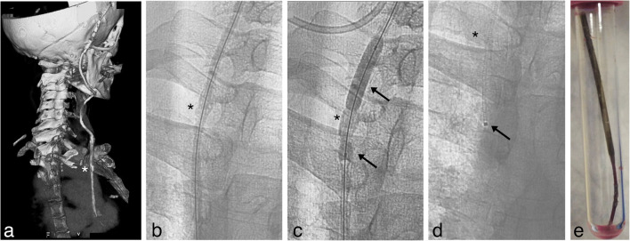
Results: We identified two patients that were implanted 28 and 40 years earlier. Both developed endocarditis with a large atrial thrombus and were successfully treated endovascularly. The successful percutaneous removal was achieved by applying, for the first time in this setting, the endoluminal dilation technique as proposed by Hong. After ventriculoatrial shunt removal and its substitution with an external drainage, both patients where successfully weaned from the need for a shunt and their infection resolved.

Conclusion: Patients carrying a ventriculoatrial shunt are now rarely seen and awareness of long-term ventriculoatrial shunt complications is decreasing. However, these complications must be recognized and treated by shunt removal. Endovascular techniques are appropriate even in the presence of overt endocarditis, atrial thrombi, and tight adherence to the endocardial wall. Moreover, weaning from shunt dependence is possible even decades after shunting.

Keywords: Endocarditis; Endovascular removal; Hydrocephalus; Right atrium; Ventriculoatrial shunt.


Conflict of interest statement

The authors declare that they have no conflict of interest.

Figures

Fig. 1
Fig. 1
Atrial condition before VAS removal. a Preoperative thoracic axial CT scan with contrast at the level of the atrium showing the VAS catheter (arrow) in patient 1. b Preoperative coronal reconstruction of the trajectory of the VAS catheter in patient 1 in the internal jugular vein, superior vena cava and atrium, radiolucency of the catheter was increased by calcium deposition in the reactive tissue surrounding the catheter, the arrow indicates the presence of macroscopic calcifications around the catheter. c Preoperative thoracic axial CT scan with contrast at the level of the atrium showing the VAS catheter in patient 2. The arrow indicates a large atrial thrombus associated with the catheter. d Axial thoracic CT scan of patient 2, the arrow indicates the VA catheter inside the right atrium. e [18F] fluoro-2-deoxy-d-glucose (FDG) PET scan image corresponding to thoracic CT scan in 1d, FDG accumulation (SUV 7.4) in close association with the position of the VAS-catheter (arrow) is visible. f Composite image demonstrating the colocalization of the FDG accumulation with the thrombus and reactive tissue surrounding the atrial catheter (arrow)
Fig. 2
Fig. 2
Removal of VAS catheter in patient 1. a Preoperative 3D CT scan reconstruction of the VAS catheter path from the valve to the right atrium, white asterisk (*) marks the site of macroscopic calcifications surrounding the catheter. b Fluoroscopy antero-posterior projection, the 0,018 inch guidewire is visible inside the VAS catheter, black asterisk (*) marks the site of the same macroscopic calcification shown in Fig. 1a. c Fluoroscopy antero-posterior projection, the VAS catheter was dilated by a 6 mm diameter Sterling balloon catheter. The calcification surrounding the VAS catheter partially restricts the dilation of the lumen compared to the adjacent segments (arrows), asterisk as in Fig. 1b. d Fluoroscopy antero-posterior projection, obtained after complete removal of the VAS catheter arrow points to an angiography catheter introduced after the VAS catheter was completely removed. e Picture of the distal portion of the VAS catheter immediately after extraction, the catheter is encrusted by calcified reactive tissue and partially deformed
Fig. 3
Fig. 3
Removal of VAS catheter in patient 2. a Fluoroscopy antero-posterior projection, a 0.018 inch guidewire is visible inside the atrial catheter (arrow), a safety wire introduced through the femoral vein (arrowhead) was placed as a guard. b Fluoroscopy antero-posterior projection, the VAS catheter was dilated by a 3.5-mm diameter Amphirion balloon catheter. Arrow and arrowhead as in Fig. 3a. c Digital subtraction antero-posterior radiogram, the VAS catheter was distally dilated by a 3.5-mm diameter Amphirion balloon catheter reaching the distal end of the VAS catheter. Arrow and arrowhead as in Fig. 3a. d Fluoroscopy antero-posterior projection, obtained after complete removal of the VAS catheter with the safety wire (arrowhead) and guidewire still in place (arrow). e Picture of the distal portion of the VAS catheter obtained immediately after extraction, the catheter is almost completely clean from thrombotic material that was present in vivo
Fig. 4
Fig. 4
Stability of ventricular system after VAS inactivation. CT scans obtained from patient 1 (a and b) and patient 2 (c and d) before surgery (a and c) and after removal of the VAS catheter from the atrium and definitive inactivation of the shunts (b and d): no appreciable increase in the diameter of the cyst and ventricles is visible. Arrows in Fig. 4a and b point to the calcified wall of the cyst
Fig. 5
Fig. 5
Plot of the probability of working VAS with time. Data for plot were derived from the literature together with the present cases (red). Median lag before VAS removal for thrombus formation and/or infection was 23 years (min 5 years, max 44.5 years, n = 23 patients) while median lag for rupture and or migration of the atrial catheter was 1.29 years (min 0.16 years, max 28 years, n = 8 patients)

References

    1. Akram Q, Saravanan D, Levy R. Valvuloplasty for tricuspid stenosis caused by a ventriculoatrial shunt. Catheter Cardiovasc Interv. 2011;77(5):722–725. doi: 10.1002/ccd.22745. - DOI - PubMed
    1. Amelot A, Bouazza S, George B, Bresson D. Causative role of infection in chronic non-thromboembolic pulmonary hypertension following ventriculo-atrial shunt. Br J Neurosurg. 2014;28(4):559–561. doi: 10.3109/02688697.2013.854311. - DOI - PubMed
    1. Arze RS, Rashid H, Morley R, Ward MK, Kerr DN. Shunt nephritis: report of two cases and review of the literature. Clin Nephrol. 1983;19(1):48–53. - PubMed
    1. Bastian D, Fessele K, Bednarski P, Göhl K. Interventional laser-assisted extraction of an infected trapped ventriculoatrial shunt based on techniques for pacemaker lead explantation. J Neurol Surg A Cent Eur Neurosurg. 2016;77(01):073–076. - PubMed
    1. Bellamy CM, Roberts DH, Ramsdale DR. Ventriculo-atrial shunt causing tricuspid endocarditis: its percutaneous removal. Int J Cardiol. 1990;28(2):260–262. doi: 10.1016/0167-5273(90)90071-C. - DOI - PubMed

Publication types

LinkOut - more resources