. 2021 Feb 8;6(3):e143958.
doi: 10.1172/jci.insight.143958.

Enhancing durability of CIS43 monoclonal antibody by Fc mutation or AAV delivery for malaria prevention

Affiliations

Enhancing durability of CIS43 monoclonal antibody by Fc mutation or AAV delivery for malaria prevention

Neville K Kisalu et al. JCI Insight. .

Abstract

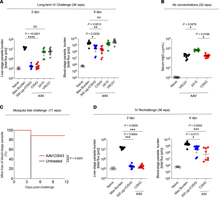
CIS43 is a potent neutralizing human mAb that targets a highly conserved "junctional" epitope in the Plasmodium falciparum (Pf) circumsporozoite protein (PfCSP). Enhancing the durability of CIS43 in vivo will be important for clinical translation. Here, 2 approaches were used to improve the durability of CIS43 in vivo while maintaining potent neutralization. First, the Fc domain was modified with the LS mutations (CIS43LS) to increase CIS43 binding affinity for the neonatal Fc receptor (FcRn). CIS43LS and CIS43 showed comparable in vivo protective efficacy. CIS43LS had 9- to 13-fold increased binding affinity for human (6.2 nM versus 54.2 nM) and rhesus (25.1 nM versus 325.8 nM) FcRn at endosomal pH 6.0 compared with CIS43. Importantly, the half-life of CIS43LS in rhesus macaques increased from 22 days to 39 days compared with CIS43. The second approach for sustaining antibody levels of CIS43 in vivo is through adeno-associated virus (AAV) expression. Mice administered once with AAV-expressing CIS43 had sustained antibody levels of approximately 300 μg/mL and mediated protection against sequential malaria challenges up to 36 weeks. Based on these data, CIS43LS has advanced to phase I clinical trials, and AAV delivery provides a potential next-generation approach for malaria prevention.

Keywords: Immunoglobulins; Infectious disease; Malaria; Skin.


Conflict of interest statement

Conflict of interest: NKK, AHI, and RAS have submitted a patent application (E-038-2017) describing CIS43 and related mutants.

Figures

Figure 1
Figure 1. Characterization of CIS43LS.
(A) Binding of CIS43LS to rPfCSP and PfCSP peptides by ELISA. CIS43, VRC01 (human anti-HIV-1 IgG1), and 317 (human antibody specific for the NANP-repeat region of PfCSP) were used as control antibodies. (B) Thermodynamic parameters and stoichiometry of binding of CIS43LS to rPfCSP by isothermal calorimetry. The KD, change in Gibbs energy (ΔG) of binding, enthalpy (ΔH), entropy contribution to Gibbs energy (−TΔS), and stoichiometry (N) are shown. Data displayed are representative of 2 independent experiments. (C) Protective effect of CIS43LS on liver burden. Following passive transfer of CIS43LS or CIS43, C57BL/6 mice were challenged i.v. with Pb-PfCSP-LUC SPZ before imaging by IVIS. Differences in liver-stage parasite burden reduction between the VRC01, CIS43, or CIS43LS group compared with the untreated group were determined using the nonparametric Kruskal-Wallis test for multiple comparisons with Dunn’s correction. P values are displayed on the panels. **P = 0.0019 (CIS43) or 0.0099 (CIS43LS). (D) Protective effect on liver burden by CIS43LS at varying concentrations (30–300 μg/mL). Data represent geometric mean with 95% CI (C and D). (E) Sterile protection by CIS43LS following infection by mosquito bites. C57BL/6 mice were challenged with infected mosquitoes following passive transfer of CIS43LS or CIS43 (300 μg per mouse). The presence of parasites was determined through Giemsa staining of blood up to 12 days following challenge. Kaplan-Meier curves, analyzed by the log rank test, show frequencies of mice free of parasites as determined by Giemsa staining of blood. Differences between CIS43LS and CIS43 as compared with untreated mice are shown (P = 0.0001). n = 5 per group (CE). (F) Apparent affinity of CIS43LS to human and rhesus FcRn at acidic pH 6.0 and physiological pH 7.4. Antibody binding curves are shown in red (raw data) and black (fitted data). The apparent affinity is displayed as KD in nM. A representative of 2 independent experiments is shown. ND, no fit could be determined.
Figure 2
Figure 2. Pharmacokinetic study in Indian rhesus macaques.
(A) Serum concentrations of CIS43LS were measured by ELISA following i.v. injection of 10 mg per kg body weight of CIS43 or CIS43LS in rhesus macaques. ****P = <0.0003. (B) The amount of mAb in skin punch homogenates from each group of CIS43- or CIS43LS-treated macaques displayed in A was quantitated over time and is shown. *P = <0.0148. Data represent the mean ± SD, and differences in mAb concentrations between both groups were determined using 2-way ANOVA (A and B). (C and D) ELISA to assess the NHP serum antibodies elicited against CIS43 or CIS43LS (antidrug antibodies, ADA). Competitive ADA ELISA (C) showing CIS43 or CIS43LS binding to rPfCSP in the presence of NHP sera before (Pre) or at the indicated time points after mAb infusion. mAb groups and animals infused with CIS43LS (12M069 and 14D017) or CIS43 (13D084 and 15C049) are indicated. Varying concentrations of mouse mAb 1-1, a CIS43 mouse anti-idiotype antibody, were used as a positive control for the competitive binding of CIS43LS or CIS43 to rPfCSP. Pre, preimmune serum collected at baseline prior to antibody infusion. OD405nm, optical density at 405 nm. Data collected at different time points from the animals that received CIS43LS (red) or CIS43 (blue) are shown; black, mAb 1-1. Direct ADA ELISA (D). CIS43LS or CIS43 was coated onto ELISA plates, and NHP sera were analyzed. Color codes are as in C. Data represent the mean ± SEM (C and D). For all panels, n = 2 per group.
Figure 3
Figure 3. Protective efficacy and expression of PfCSP-AAV–induced mAbs in C57BL/6 mice.
(A) Sterile protection by mosquito bite 3 weeks following intramuscular administration of 1 × 10¹¹ genome copies (GC) AAV encoding CIS43, 2A10, or VRC01. C57BL/6 mice were challenged by mosquito bites 3 weeks following AAV administration of the indicated mAb. As a control, 300 μg-rCIS43 was administered 2 hours prior to challenge. Kaplan-Meier curves, analyzed using the log-rank test, show the frequencies of mice free of parasites. Differences between CIS43-AAV–administered or 300 μg rCIS43–treated mice and untreated mice are shown (P = 0.0001). wpa, weeks post-AAV administration; 300 μg-rCIS43, mice that passively received the protective dose of 300 μg of CIS43 at the time of challenge. n = 10 per group; 2A10-AAV, n = 8. (B) Protective effect of PfCSP-AAV expressed mAbs on liver burden 8 weeks following AAV administration. AAV-treated C57BL/6 albino mice were challenged i.v. with Pb-PfCSP-LUC SPZ and imaged by IVIS. Data represent the geometric mean with 95% CI. For liver burden (2 dpc), *P = 0.0159 and ***P = 0.0001; for parasitemia (6 dpc), *P = 0.0102 and ***P = 0.0005. Max burden, naive infected mice; 8 wpa, 8 weeks post-AAV administration; dpc, days postchallenge; hIgG; human IgG1. CIS43 and VRC01, n = 10 per group; 2A10, n = 8. (CF) Serum hIgG (C) and PfCSP-specific mAb (D) levels, and skin hIgG (E) and PfCSP-specific mAb (F) levels from mice shown in B were measured by ELISA 8 wpa. Differences in antibody concentration, parasite liver burden, or parasitemia between each group and the untreated group were determined using the Kruskal-Wallis test for multiple comparisons with Dunn’s correction. P values are shown on panels. Means ± SD are displayed (CF).
Figure 4
Figure 4. Long-term expression and protective capacity of AAV-induced mAbs.
(A) C57BL/6 albino mice were challenged i.v. with Pb-PfCSP-LUC SPZ 36 weeks after a single administration of AAV encoding CIS43, 2A10, or VRC01. As a control, rCIS43 (300 μg) administered 2 hours prior to challenge. For all groups, n = 10; 2A10-AAV, n = 8. *P = 0.0235; **P = 0.0013; ****P ≤ 0.0001. (B) Serum hIgG levels of AAV-injected mice shown in A were measured by ELISA 32 weeks after AAV administration prior rechallenge (36 weeks). P values are shown on the panel. Data represent the mean ± SEM. (C) AAV-treated C57BL/6 mice previously protected at 3 weeks after CIS43-AAV administration (Figure 3A) were rechallenged by mosquito bites 11 weeks following the AAV administration. Kaplan-Meier curves, analyzed using the log-rank test, show the frequencies of mice free of parasites as determined through Giemsa staining of blood up to 12 days following challenge. Differences between CIS43-AAV–administered mice and untreated mice are shown (****P = 0.0001). Untreated mice, n = 7; CIS43-AAV, n = 9. (D) C57BL/6 albino mice previously protected when challenged 8 weeks after AAV administration (Figure 3B) were rechallenged i.v. with Pb-PfCSP-LUC SPZ at 36 weeks. Liver burden (left) and parasitemia (right) are shown. P values are displayed on the panels. CIS43-AAV, n = 8; 300 μg-rCIS43, n = 10. Differences in parasite liver burden, parasitemia, or serum antibody concentration between groups were determined using the Kruskal-Wallis test for multiple comparisons with Dunn’s correction. Data represent the geometric mean with 95% CI (A and D).

References

    1. Mbacham WF, et al. Current situation of malaria in Africa. Methods Mol Biol. 2019;2013:29–44. doi: 10.1007/978-1-4939-9550-9_2. - DOI - PubMed
    1. WHO. World Malaria Report. Wold Health Organization; 2016.
    1. Olotu A, et al. Four-year efficacy of RTS,S/AS01E and its interaction with malaria exposure. N Engl J Med. 2013;368(12):1111–1120. doi: 10.1056/NEJMoa1207564. - DOI - PMC - PubMed
    1. Olotu A, et al. Seven-year efficacy of RTS,S/AS01 malaria vaccine among young African children. N Engl J Med. 2016;374(26):2519–2529. doi: 10.1056/NEJMoa1515257. - DOI - PMC - PubMed
    1. Epstein JE, et al. Protection against Plasmodium falciparum malaria by PfSPZ Vaccine. JCI Insight. 2017;2(1):e89154. - PMC - PubMed

Publication types

MeSH terms

LinkOut - more resources