. 2021 Feb 15;131(4):e141335.
doi: 10.1172/JCI141335.

Regulation and targeting of androgen receptor nuclear localization in castration-resistant prostate cancer

Affiliations

Regulation and targeting of androgen receptor nuclear localization in castration-resistant prostate cancer

Shidong Lv et al. J Clin Invest. .

Abstract

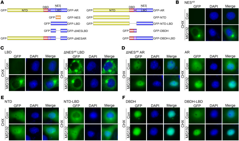
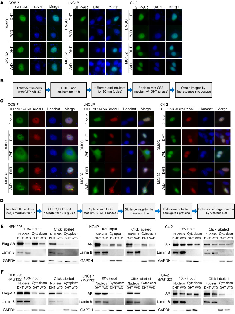
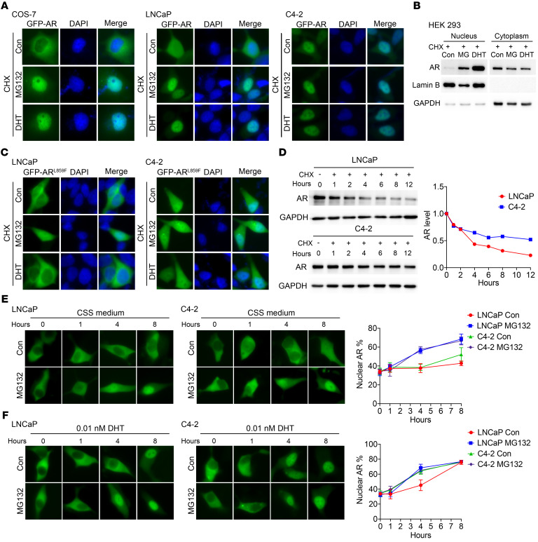
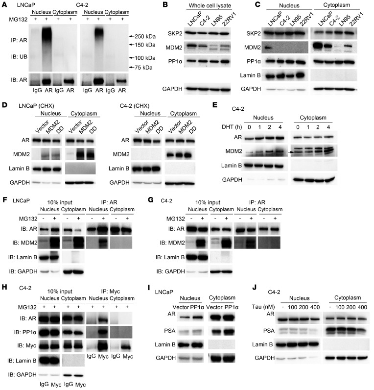
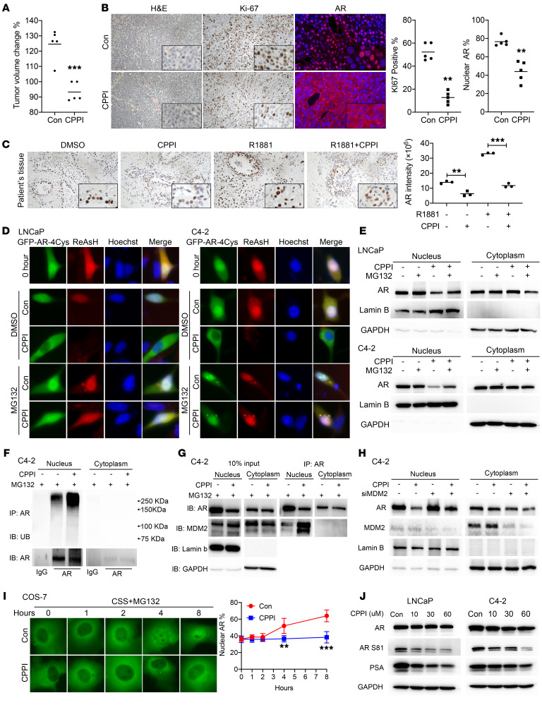
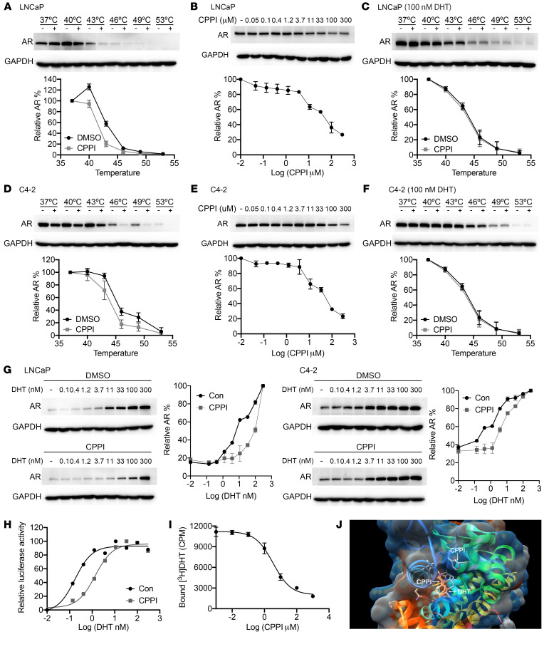
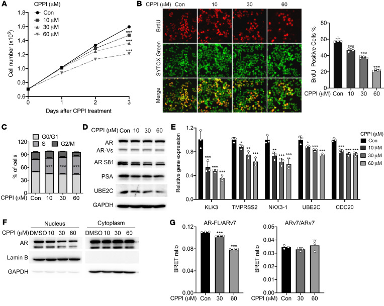
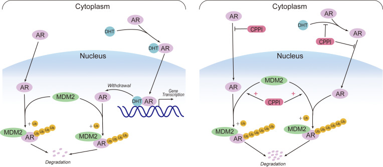
Nuclear localization of the androgen receptor (AR) is necessary for its activation as a transcription factor. Defining the mechanisms regulating AR nuclear localization in androgen-sensitive cells and how these mechanisms are dysregulated in castration-resistant prostate cancer (CRPC) cells is fundamentally important and clinically relevant. According to the classical model of AR intracellular trafficking, androgens induce AR nuclear import and androgen withdrawal causes AR nuclear export. The present study has led to an updated model that AR could be imported in the absence of androgens, ubiquitinated, and degraded in the nucleus. Androgen withdrawal caused nuclear AR degradation, but not export. In comparison with their parental androgen-sensitive LNCaP prostate cancer cells, castration-resistant C4-2 cells exhibited reduced nuclear AR polyubiquitination and increased nuclear AR level. We previously identified 3-(4-chlorophenyl)-6,7-dihydro-5H-pyrrolo[1,2-a]imidazole (CPPI) in a high-throughput screen for its inhibition of androgen-independent AR nuclear localization in CRPC cells. The current study shows that CPPI is a competitive AR antagonist capable of enhancing AR interaction with its E3 ligase MDM2 and degradation of AR in the nuclei of CRPC cells. Also, CPPI blocked androgen-independent AR nuclear import. Overall, these findings suggest the feasibility of targeting androgen-independent AR nuclear import and stabilization, two necessary steps leading to AR nuclear localization and activation in CRPC cells, with small molecule inhibitors.

Keywords: Drug therapy; Oncology; Prostate cancer; Therapeutics.


Conflict of interest statement

Conflict of interest: JBN, PW, and Z Wang are listed as co-inventors on a US patent held by the University of Pittsburgh (US 10004730, “Small molecules targeting androgen receptor nuclear localization and/or level in prostate cancer”).

Figures

Figure 1
Figure 1. Effect of MG132 on subcellular localization of GFP-tagged AR and AR deletion constructs.
(A) Diagrams of GFP-tagged AR and AR deletion constructs: GFP-AR, GFP-NESAR, GFP-LBD, GFP-ΔNESARLBD, GFP-ΔNESARAR, GFP-NTD, GFP-NTD-LBD, GFP-DBDH, and GFP-DBDH-LBD. (BF) Representative fluorescent images of transfected GFP-NESAR (B), GFP-LBD, GFP-ΔNESARLBD (C), GFP-ΔNESARAR, GFP-AR (D), GFP-NTD, GFP-NTD-LBD (E), GFP-DBDH, and GFP-DBDH-LBD (F) in COS-7 cells 24 hours after treatment with DMSO, CHX, and/or MG132. Con, control. Data represent 1 of 2 independent experiments with consistent results. Original magnification, ×40.
Figure 2
Figure 2. Imported nuclear AR is degraded and not exported following DHT withdrawal.
(A) Representative fluorescent images of transfected GFP-AR in COS-7, LNCaP, or C4-2 cells treated with 10 nM DHT followed by DHT withdrawal (W/D) and CHX in presence or absence of MG132. (B) Schematic of ReAsH pulse-chase experiments. (C) Representative fluorescent images of ReAsH pulse-labeled GFP-AR-4Cys. Green signal represented total GFP-AR-4Cys protein, whereas red signal was derived from ReAsH-labeled GFP-AR-4Cys protein. Original magnification, ×40.(D) Schematic of Click pulse-chase analysis. (E and F) Western blot detection of the pulsed labeled protein in cells cultured in the absence (E) or presence (F) of MG132, with (DHT) or without (W/D) DHT. Data represent 1 of at least 2 independent experiments with consistent results.
Figure 3
Figure 3. Unliganded AR can be imported into the nucleus in the presence of MG132.
(A) Representative fluorescent images of GFP-AR in COS-7, LNCaP, and C4-2 cells 24 hours after treatment with MG132 or DHT in CSS medium containing CHX. (B) Western blot analysis of nuclear and cytoplasmic extracts of transiently transfected Flag-AR in HEK293 cells after treatment with MG132 or DHT in CSS medium containing CHX. (C) Representative fluorescent images of GFP-ARL859F after treatment with MG132 or DHT for 24 hours in LNCaP or C4-2 cells in CSS medium containing CHX. (D) Effect of CHX on the protein levels of endogenous AR at indicated time points in LNCaP or C4-2 cells. (E and F) GFP-AR–transfected LNCaP and C4-2 cells were pretreated with CPPI for 24 hours and then replaced with fresh CSS medium without CPPI, but containing no DHT (E) (n = 6) or 0.01 nM DHT (F) (n = 6) with or without MG132. The GFP-AR images were detected at indicated time points after the medium replacement. Nuclear GFP-AR quantification data are shown at right. Quantitative data are presented as mean ± SD, and all data represent 1 of at least 2 independent experiments with consistent results. Original magnification, ×40.
Figure 4
Figure 4. AR is polyubiquitinated and associated with E3 ligase MDM2 in the nuclei.
(A) Western blot analysis of ubiquitin (UB) and AR in LNCaP and C4-2 cells in the presence of MG132. (B and C) Western blot analysis of AR, SKP2, MDM2, and PP1α in whole-cell lysate (B) or nucleocytoplasmic fractions (C). (D) LNCaP and C4-2 cells were transfected with MDM2 or MDM2S166D, S186D (DD) expression vector. Expression levels of nuclear and cytoplasmic AR in presence of CHX were detected through Western blot. (E) C4-2 cells were cultured in CSS medium for 48 hours, and the time course of AR and MDM2 localization after DHT treatment was detected through Western blot. (F and G) Nuclear and cytoplasmic extracts were prepared from the MG132-treated LNCaP (F) and C4-2 (G) for IP with anti-AR antibody. Immunoblotting was performed using indicated antibodies. (H) IP was performed in myc-MDM2–transfected C4-2 cells with anti-myc antibody. IB was conducted with indicated antibodies. (I) Western blot analysis of protein levels of AR in LNCaP cells after PP1α transfection in CSS medium. (J) Effect of tautomycin (Tau) on expression levels of nuclear and cytoplasm AR was detected through Western blot in C4-2 cells. Data represent 1 of at least 2 independent experiments with consistent results.
Figure 5
Figure 5. CPPI enhanced nuclear AR polyubiquitination and degradation, increased AR association with MDM2 in the nucleus, and inhibited AR nuclear import.
(A) Quantification of tumor volume changes after CPPI treatment (50 mg/kg/d) in C4-2 xenograft tumors (n = 5). (B) Representative images of H&E, Ki-67 immunostaining, and AR immunofluorescent staining in C4-2 xenograft tumors treated with CPPI or vehicle (n = 3). (C) Patient-derived explants were treated with CPPI. Effects of CPPI on AR expression with representative sections are shown. (D) Representative fluorescent images of total (green signal) and ReAsH pulse-labeled (red signal) GFP-AR-4Cys in response to CPPI (30 μM) in LNCaP or C4-2 cells with or without MG132. (E) Western blot analysis of endogenous AR in nuclear and cytoplasmic extracts of LNCaP and C4-2 cells after CPPI treatment with or without MG132 as in (C). (F) Western blot analysis of ubiquitin and AR in C4-2 cells treated with or without CPPI for 24 hours in the presence of MG132. (G) IP samples shown in E were also analyzed by Western blot with additional indicated antibodies. (H) Western blot analysis of AR expression in MDM2 knockdown or control C4-2 cells after CPPI treatment. (I) Time course of GFP-AR localization in transfected COS-7 cells treated with or without CPPI in the presence of MG132 in CSS medium. Nuclear GFP-AR quantification data are shown at right (n = 6). (J) AR, AR S81, and PSA were detected by Western blot in LNCaP and C4-2 cells treated with CPPI. Quantitative data are presented as mean ± SD, and all data represent 1 of at least 2 independent experiments with consistent results. Unpaired t test (I) was used to determine statistical significance. **P < 0.01; ***P < 0.001. Original magnification, ×40.
Figure 6
Figure 6. CPPI is an AR competitive inhibitor.
(A) Western blots showing thermostable AR following indicated heat shocks in the presence (+) or absence (−) of 50 μM CPPI in LNCaP cells (n = 2). (B) ITDRFCETSA experiments to determine the potency of CPPI target engagement in LNCaP cells (n = 2). (C) Western blots showing thermostable AR following indicated heat shocks in the presence (+) or absence (−) of 50 μM CPPI with 100 nM DHT in LNCaP cells (n = 2). (DF) Western blots showing thermostable AR (D) (n = 2), the potency of CPPI target engagement (E) (n = 2),and the thermostable AR with 100 nM DHT (F) (n = 2) in C4-2 cells. (G) ITDRFCETSA experiments performed for DHT in the presence of 50 μM CPPI in LNCaP and C4-2 cells (n = 2). (H) C4-2-PSA-rl cells were treated with a gradient DHT dose with or without 50 μM CPPI. Firefly luciferase values were determined and normalized to Renilla (n = 3). (I) C4-2 cells were incubated with 1 nM [3H] DHT and the indicated amount of CPPI. The retained [3H] DHT in C4-2 cells was counted (n = 3). (J) Illustration of in silico predicted CPPI-binding sites in the LBD of AR. Quantitative data are presented as mean ± SD, and all data represent 1 of 2 independent experiments with consistent results.
Figure 7
Figure 7. CPPI inhibited LNCaP95 cell proliferation and full-length AR interaction with ARv7.
(A) LNCaP95 cell proliferation following treatment with indicated concentrations of CPPI for 1, 2, and 3 days (n = 3). (B) BrdU incorporation in LNCaP95 cells treated with CPPI. Right panel shows percentages of the LNCaP95 cells stained with BrdU (n = 4). Original magnification, ×40. (C) Cell-cycle analysis of LNCaP95 cells treated with CPPI (n = 3). (D) Western blot of AR, AR S81, PSA, and UBE2C in LNCaP95 cells treated with CPPI. GAPDH was probed as loading control. (E) qPCR analysis of AR target genes (KLK3, TMPRSS2, and NKX3-1) and ARV target genes (UBE2C and CDC20) in LNCaP95 cells treated with CPPI (n = 3). (F) Western blot analysis of AR and ARv7 in the nuclear and cytoplasmic extracts of LNCaP95 cells treated with CPPI. (G) BRET assay of AR-FL and ARv7 interaction following CPPI treatment (n = 3). Quantitative data are presented as mean ± SEM, and all data represent 1 of at least 2 independent experiments with consistent results. Unpaired t test (A) or 1-way ANOVA with Dunnett’s multiple-comparison post test (B, C, E, and G) was used to determine statistical significance. *P < 0.05; **P < 0.01; ***P < 0.001.
Figure 8
Figure 8. An updated model for AR intracellular trafficking and the mechanisms of CPPI targeting nuclear AR.
AR can be imported in the absence of androgens, and the imported AR will be efficiently polyubiquitinated and degraded via proteasomes in the nucleus. DHT binding enhances AR nuclear import and inhibits AR degradation in the nucleus. Once AR is in the nucleus, it will not be exported. After DHT withdrawal, unliganded AR will undergo degradation in the nucleus immediately (left). In CRPC cells, the increased nuclear AR stability will lead to AR nuclear localization. CPPI, a pyrroloimidazole, can block AR ligand binding, inhibit both androgen-induced and androgen-independent AR import, and act as a nuclear AR degrader in CRPC cells (right).

References

    1. Siegel RL, et al. Cancer statistics, 2020. CA Cancer J Clin. 2020;70(1):7–30. doi: 10.3322/caac.21590. - DOI - PubMed
    1. de Bono JS, et al. Abiraterone and increased survival in metastatic prostate cancer. N Engl J Med. 2011;364(21):1995–2005. doi: 10.1056/NEJMoa1014618. - DOI - PMC - PubMed
    1. Ryan CJ, et al. Abiraterone in metastatic prostate cancer without previous chemotherapy. N Engl J Med. 2013;368(2):138–148. doi: 10.1056/NEJMoa1209096. - DOI - PMC - PubMed
    1. Salem M, Garcia JA. Abiraterone acetate, a novel adrenal inhibitor in metastatic castration-resistant prostate cancer. Curr Oncol Rep. 2011;13(2):92–96. doi: 10.1007/s11912-011-0153-4. - DOI - PubMed
    1. Tran C, et al. Development of a second-generation antiandrogen for treatment of advanced prostate cancer. Science. 2009;324(5928):787–790. doi: 10.1126/science.1168175. - DOI - PMC - PubMed

Publication types

MeSH terms

Substances