. 2020 Dec 17;15(12):e0241148.
doi: 10.1371/journal.pone.0241148. eCollection 2020.

Transcriptomic and proteomic intra-tumor heterogeneity of colorectal cancer varies depending on tumor location within the colorectum

Affiliations

Transcriptomic and proteomic intra-tumor heterogeneity of colorectal cancer varies depending on tumor location within the colorectum

Sigrid Salling Árnadóttir et al. PLoS One. .

Abstract

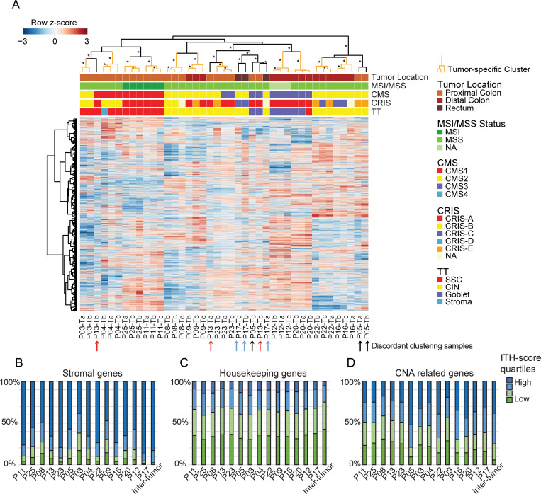
Background: Intra-tumor heterogeneity (ITH) of colorectal cancer (CRC) complicates molecular tumor classification, such as transcriptional subtyping. Differences in cellular states, biopsy cell composition, and tumor microenvironment may all lead to ITH. Here we analyze ITH at the transcriptomic and proteomic levels to ascertain whether subtype discordance between multiregional biopsies reflects relevant biological ITH or lack of classifier robustness. Further, we study the impact of tumor location on ITH.

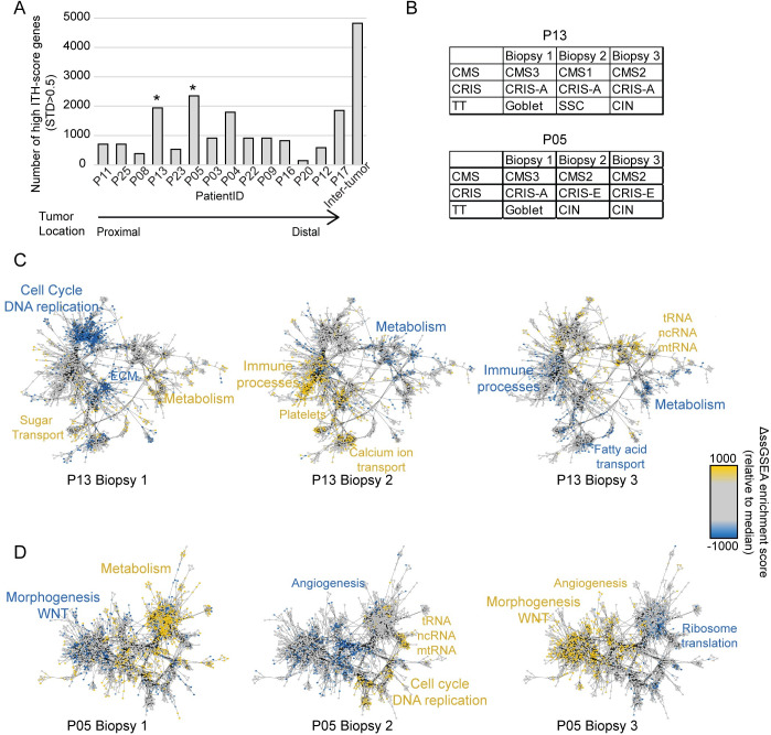
Methods: Multiregional biopsies from stage II and III CRC tumors were analyzed by RNA sequencing (41 biopsies, 14 tumors) and multiplex immune protein analysis (89 biopsies, 29 tumors). CRC subtyping was performed using consensus molecular subtypes (CMS), CRC intrinsic subtypes (CRIS), and TUMOR types. ITH-scores and network maps were defined to determine the origin of heterogeneity. A validation cohort was used with one biopsy per tumor (162 tumors).

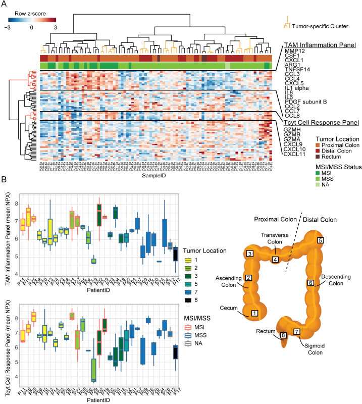
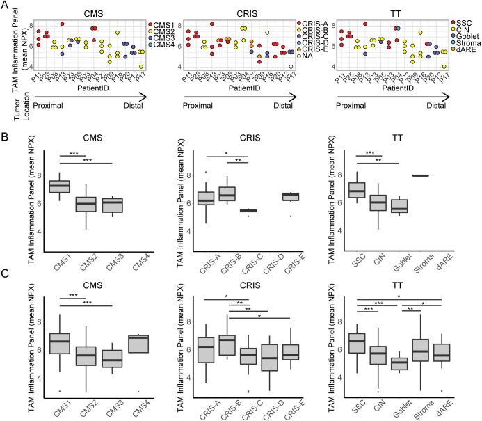
Results: Overall, inter-tumor transcriptional variation exceeded ITH, and subtyping calls were frequently concordant between multiregional biopsies. Still, some tumors had high transcriptional ITH and were classified discordantly. Subtyping of proximal MSS tumors were discordant for 50% of the tumors, this ITH was related to differences in the microenvironment. Subtyping of distal MSS tumors were less discordant, here the ITH was more cancer-cell related. The subtype discordancy reflected actual molecular ITH within the tumors. The relevance of the subtypes was reflected at protein level where several inflammation markers were significantly increased in immune related transcriptional subtypes, which was verified in an independent cohort (Wilcoxon rank sum test; p<0.05). Unsupervised hierarchical clustering of the protein data identified large ITH at protein level; as the multiregional biopsies clustered together for only 9 out of 29 tumors.

Conclusion: Our transcriptomic and proteomic analyses show that the tumor location along the colorectum influence the ITH of CRC, which again influence the concordance of subtyping.


Conflict of interest statement

The authors have declared that no competing interests exist.

Figures

Fig 1
Fig 1. Tumor location influences ITH of transcriptional CRC subtyping.
(A) All biopsies were classified using three different classifiers; CMS, CRIS and TT. Each circle depicts a tumor, and each piece a biopsy. All tumors are presented three times, one for each classifier. * denotes CMS subtype calls that were only called, when using the SSP.nearest method, in contrast the SSP.predicted method left these samples unclassified. CRIS classifications with multiple colors are due to uncertain subtype calls. (B) Discordant or concordant subtype calls within tumors, for all tumors, proximal tumor, and distal tumors (grey = discordant subtype calls within a tumor, blue = concordant subtype calls). (C) Caleydo Stratomex plots of the distribution and correlation between subtypes called for each biopsy. Top = highlighted for proximal tumors, bottom = highlighted for distal tumors. (CMS = consensus molecular subtypes, CRIS = CRC intrinsic subtypes, TT = tumor types).
Fig 2
Fig 2. Transcriptional ITH.
(A) Transcriptional heatmap of protein coding genes. For 11/14 tumors, the multiregional biopsies cluster in tumor-specific clusters (marked with orange). Biopsies from the remaining 3 tumors are combined in clusters (marked in black). The clusters indicated with asterisk (*) were statistical significant as evaluated by bootstrapping (Approximately Unbiased (AU) values ≥95). Arrows below the heatmap indicate discordant clustering biopsies (black: P05, red: P13, blue: P17). Annotations above the heatmap illustrates tumor location, MSI/MSS status, and subtype calls for all three classifiers. The majority of samples cluster according to their subtype combination. (B-D) Distribution of ITH-score for three gene panels, the four colors represent quartiles ranging from low to high ITH-score. Patient specific ITH-scores are calculated as standard deviation for each gene between all biopsies from each tumor. Inter-tumor denotes variation between tumors and is calculated as the STD between all biopsies from all tumors. (B) A panel of stromal genes (n = 618) are enriched for high ITH-score genes. (C) A panel of housekeeping genes (n = 3415) are more stable both within and between tumors. (D) Transcripts related to common copy number alterations (CNAs) are equally distributed in all four categories for most patients (n = 831). P05 have a larger fraction of high ITH-score genes related to CNAs.
Fig 3
Fig 3. ITH of molecular pathways.
(A) Barplot with quantification of number of genes with high ITH-score (STD > 0.5) for each tumor and all tumors combined. * denotes tumors with the highest amount of high ITH-score genes, which are included in B-D. (B) Subtyping results for each biopsy from tumor P13 and P05. (C-D) Tumor-specific network maps for two tumors illustrating the 5000 ssGSEA terms with the highest ITH for tumor P13 in (C) and tumor P05 in (D). Yellow dots/font indicates mechanisms that are upregulated in the sample compared to the other samples from the same tumor, while blue indicates downregulated mechanisms.
Fig 4
Fig 4. ITH of immune response on protein level.
(A) Heatmap of protein levels based on the immuno-oncology panel. All columns represent a sample; biopsies in tumor-specific clusters are marked with orange (9/29 tumors). Annotations indicate tumor location and MSI/MSS status. Row-side trees marked with red represent a TAM Inflammation Panel and Tcyt Cell Response Panel. (B) Boxplots showing calculated sample-means for TAM Inflammation panel (top) and the Tcyt cell panel (bottom) for each tumor. Fill colors (1–8) indicate tumor location as illustrated in the schematic figure of the colon and rectum. Red/blue border colors indicate MSI/MSS status. Each bar illustrates results from all biopsies from each tumor. (TAM = tumor associated macrophage, Tcyt = cytotoxic T cell).
Fig 5
Fig 5. TAM Inflammation on protein level varies between subtypes.
(A) Dotplots showing TAM inflammation panel (mean NPX) protein level for multiregional biopsies from each tumors (n = 14) with all three classifiers (CMS, CRIS, and TT). Colors indicate transcriptional subtype. Proximal to distal tumor location is indicated by an arrow. (B) Boxplots for each classifier (CMS, CRIS, TT), showing protein inflammation panel mean for all samples (n = 41) grouped based on transcriptional subtype. (C) Boxplots for each classifier, showing protein inflammation panel mean for samples from a validation cohort (n = 162), grouped based on transcriptional subtype. (* = p<0.05; ** = p<0.01; *** = p<0.001, Wilcoxon rank sum test).

References

    1. Bray F, Ferlay J, Soerjomataram I, Siegel RL, Torre LA, Jemal A. Global cancer statistics 2018: GLOBOCAN estimates of incidence and mortality worldwide for 36 cancers in 185 countries. CA: a cancer journal for clinicians. 2018;68(6):394–424. - PubMed
    1. Puppa G, Sonzogni A, Colombari R, Pelosi G. TNM staging system of colorectal carcinoma: a critical appraisal of challenging issues. Arch Pathol Lab Med. 2010;134(6):837–52. 10.1043/1543-2165-134.6.837 - DOI - PubMed
    1. Guinney J, Dienstmann R, Wang X, de Reynies A, Schlicker A, Soneson C, et al. The consensus molecular subtypes of colorectal cancer. Nature medicine. 2015;advance online publication. 10.1038/nm.3967 - DOI - PMC - PubMed
    1. Bramsen JB, Rasmussen MH, Ongen H, Mattesen TB, Ørntoft M-BW, Árnadóttir SS, et al. Molecular-Subtype-Specific Biomarkers Improve Prediction of Prognosis in Colorectal Cancer. Cell reports. 2017;19(6):1268–80. 10.1016/j.celrep.2017.04.045 - DOI - PubMed
    1. Isella C, Brundu F, Bellomo SE, Galimi F, Zanella E, Porporato R, et al. Selective analysis of cancer-cell intrinsic transcriptional traits defines novel clinically relevant subtypes of colorectal cancer. Nat Commun. 2017;8:15107 10.1038/ncomms15107 - DOI - PMC - PubMed

Publication types

MeSH terms