

Lipid alterations in human frontal cortex in ALS-FTLD-TDP43 proteinopathy spectrum are partly related to peroxisome impairment
- PMID: 33332650
- PMCID: PMC8248144
- DOI: 10.1111/nan.12681
Lipid alterations in human frontal cortex in ALS-FTLD-TDP43 proteinopathy spectrum are partly related to peroxisome impairment
Abstract
Aim: Peroxisomes play a key role in lipid metabolism, and peroxisome defects have been associated with neurodegenerative diseases such as X-adrenoleukodystrophy and Alzheimer's disease. This study aims to elucidate the contribution of peroxisomes in lipid alterations of area 8 of the frontal cortex in the spectrum of TDP43-proteinopathies. Cases of frontotemporal lobar degeneration-TDP43 (FTLD-TDP), manifested as sporadic (sFTLD-TDP) or linked to mutations in various genes including expansions of the non-coding region of C9ORF72 (c9FTLD), and of sporadic amyotrophic lateral sclerosis (sALS) as the most common TDP43 proteinopathies, were analysed.
Methods: We used transcriptomics and lipidomics methods to define the steady-state levels of gene expression and lipid profiles.
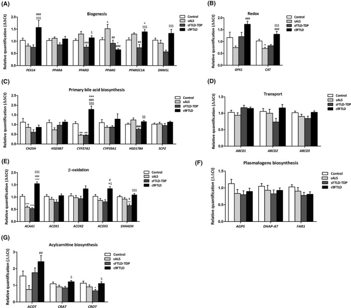
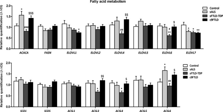
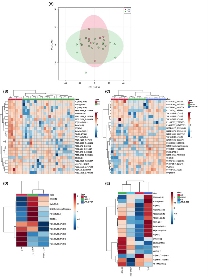
Results: Our results show alterations in gene expression of some components of peroxisomes and related lipid pathways in frontal cortex area 8 in sALS, sFTLD-TDP and c9FTLD. Additionally, we identify a lipidomic pattern associated with the ALS-FTLD-TDP43 proteinopathy spectrum, notably characterised by down-regulation of ether lipids and acylcarnitine among other lipid species, as well as alterations in the lipidome of each phenotype of TDP43 proteinopathy, which reveals commonalities and disease-dependent differences in lipid composition.
Conclusion: Globally, lipid alterations in the human frontal cortex of the ALS-FTLD-TDP43 proteinopathy spectrum, which involve cell membrane composition and signalling, vulnerability against cellular stress and possible glucose metabolism, are partly related to peroxisome impairment.
Keywords: fatty acid profiling; human frontal cortex; lipidomics; peroxisomes; plasmalogens; transcriptomics.
© 2020 The Authors. Neuropathology and Applied Neurobiology published by John Wiley & Sons Ltd on behalf of British Neuropathological Society.
Conflict of interest statement
The authors declare that the research was conducted in the absence of any commercial or financial relationships that could be construed as potential conflicts of interest.
Figures
References
-
- De Duve C, Baudhuin P. Peroxisomes (microbodies and related particles). Physiol Rev. 1996;46:323‐327. - PubMed
-
- Pedersen JI, Eggertsen G, Hellman U, Andersson U, Björkhem I. Molecular cloning and expression of cDNA encoding 3alpha, 7alpha,12alpha‐trihydroxy‐5beta‐chole stanoyl‐CoA oxidase from rabbit liver. J Biol Chem. 1997;272:18481‐18489. - PubMed
-
- Wanders RJ, Waterham HR. Biochemistry of mammalian peroxisomes revisited. Annu Rev Biochem. 2006;75:295‐332. - PubMed
-
- Islinger M, Cardoso MJ, Schrader M. Be different—the diversity of peroxisomes in the animal kingdom. Biochim Biophys Acta. 2010;1803:881‐887. - PubMed
-
- Wanders RJ. Peroxisomes in human health and disease: metabolic pathways, metabolite transport, interplay with other organelles and signal transduction. Subcell Biochem. 2013;69:23‐44. - PubMed
Publication types
MeSH terms
LinkOut - more resources
Full Text Sources
Other Literature Sources
Medical
Miscellaneous
