. 2020 Dec;91(1):e81.
doi: 10.1002/cpph.81.

Targeted Protein Degradation Phenotypic Studies Using HaloTag CRISPR/Cas9 Endogenous Tagging Coupled with HaloPROTAC3

Affiliations

Targeted Protein Degradation Phenotypic Studies Using HaloTag CRISPR/Cas9 Endogenous Tagging Coupled with HaloPROTAC3

Elizabeth A Caine et al. Curr Protoc Pharmacol. 2020 Dec.

Abstract

To assess the role of a protein, protein loss phenotypic studies can be used, most commonly through mutagenesis RNAi or CRISPR knockout. Such studies have been critical for the understanding of protein function and the identification of putative therapeutic targets for numerous human disease states. However, these methodological approaches present challenges because they are not easily reversible, and if an essential gene is targeted, an associated loss of cell viability can potentially hinder further studies. Here we present a reversible and conditional live-cell knockout strategy that is applicable to numerous proteins. This modular protein-tagging approach regulates target loss at the protein, rather than the genomic, level through the use of HaloPROTAC3, which specifically degrades HaloTag fusion proteins via recruitment of the VHL E3 ligase component. To enable HaloTag-mediated degradation of endogenous proteins, we provide protocols for HaloTag genomic insertion at the protein N or C terminus via CRISPR/Cas9 and use of HaloTag fluorescent ligands to enrich edited cells via Fluorescence-Activated Cell Sorting (FACS). Using these approaches, endogenous HaloTag fusion proteins present in various subcellular locations can be degraded by HaloPROTAC3. As detecting the degradation of endogenous targets is challenging, the 11-amino-acid peptide tag HiBiT is added to the HaloTag fusion to allows the sensitive luminescence detection of HaloTag fusion levels without the use of antibodies. Lastly, we demonstrate, through comparison of HaloPROTAC3 degradation with that of another fusion tag PROTAC, dTAG-13, that HaloPROTAC3 has a faster degradation rate and similar extent of degradation. © 2020 The Authors. Basic Protocol 1: CRISPR/Cas9 insertion of HaloTag or HiBiT-HaloTag Basic Protocol 2: HaloPROTAC3 degradation of endogenous HaloTag fusions.

Keywords: CRISPR; HaloPROTAC3; HaloTag; HiBiT; PROTAC; VHL; phenotype; targeted protein degradation.


Figures

Figure 1
Figure 1
Schematic of HaloPROTAC3 degradation of HaloTag protein fusions in live cells. First, CRISPR/Cas9 technology is used to insert HaloTag or HiBiT‐HaloTag into the genomic locus of the target protein using a dsDNA donor plasmid and Cas9‐crRNA complex (1). After the HaloTag fusion is expressed, cells are treated with HaloPROTAC3(2). HaloPROTAC3 induces a ternary complex between the VHL E3 ligase component and HaloTag protein fusion, resulting degradation of the HaloTag target protein via the ubiquitin‐proteasomal pathway (3).
Figure 2
Figure 2
Efficiency of multiple crRNAs used to insert HaloTag‐HiBiT to the C terminus of the endogenous EPOP protein was tested using a Nano‐Glo HiBiT Lytic Detection Assay. Analyzed data from raw RLUs shows the ratio of the luminescence from each CRISPR‐edited pool of cells over background luminescence from HEK293 parent cells (A). crRNA 1 has the highest signal/background ratio, correlating to the highest HiBiT‐HaloTag insertion frequency. Insertion efficiency of large tags with CRISPR/Cas9 editing is low. The fluorescent JF646 HaloTag ligand allows HaloTag‐positive cells to be sorted from negative cells, enriching the edited population (B). Labeled parental HEK293 cells (blue) were used to determine negative cells. The β‐catenin‐HaloTag cells (red) contained a large HEK293 parent peak and a small JF646‐HaloTag‐positive peak that represented ∼6% of the total live cell population.
Figure 3
Figure 3
Endpoint analysis of a homozygous β‐catenin‐HaloTag‐HiBiT endogenous protein fusion in HEK293 cells that stably express LgBiT after degradation with HaloPROTAC3. Cells contained the endogenous β‐catenin‐HaloTag‐HiBiT protein fusion were treated with HaloPROTAC3 for 3 or 24 hr, and degradation was detected with an endpoint HiBiT lytic assay (A). HaloPROTAC3 caused rapid reduction in the amount of tagged protein in the cell, which was visualized through luminescence imaging on an Olympus LV200 microscope (B). Phenotypic characterization was performed on cells that were treated with HaloPROTAC3. Cells containing the endogenous β‐catenin‐HaloTag‐HiBiT protein fusion were transfected with a TCF firefly luciferase reporter before being treating with mWnt3a. HaloPROTAC3 was used to degrade the beta‐catenin‐HaloTag‐HiBiT protein fusion, and after 24 hr a lytic dual luciferase assay was performed to measure firefly luciferase expressed from the TCF reporter (C) and β‐catenin HiBiT luminescence (D). TCF expression was not induced in cells that were treated with HaloPROTAC3 because β‐catenin‐HaloTag‐HiBiT protein fusions were degraded and could not enter the nucleus upon treatment with mWnt3a.
Figure 4
Figure 4
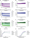
Kinetic degradation profiles of endogenous nuclear, mitochondrial membrane, and cytoplasmic HiBiT‐HaloTag protein fusions after treatment with HaloPROTAC3. Cells were treated with a series of dilutions of HaloPROTAC3 (A, C, E) or ent‐HaloPROTAC3 (B, D, F), and degradation was followed by luminescence detection in live cells through a kinetic degradation assay for 24 hr. An N‐terminal HiBiT‐HaloTag‐nuclear endogenous protein fusion in HEK293 cells that stably express LgBiT, a C‐terminal HaloTag‐HiBiT mitochondrial membrane endogenous protein fusion in parent HEK293 cells with LgBiT introduced by transient transfection, and a C‐terminal HaloTag‐HiBiT‐cytoplasmic protein fusion in HEK293 cells that stably express LgBiT were used, respectively. No degradation was detected in the ent‐HaloPROTAC3‐treated samples, confirming that the protein loss is due to a PROTAC‐mediated mechanism. (G) Rapid degradation rates were observed in all samples, with >50% degradation occurring in the first 3 hr. Around 80% degradation was achieved with each HiBiT‐HaloTag protein fusion regardless of cellular location (H). DC50 values were 8.1 nM for the mitochondrial membrane and nuclear protein and 18.6 nM for the cytoplasmic protein.
Figure 5
Figure 5
Comparison between a HaloTag insertion with HaloPROTAC3 degradation and an FKBP12F36V insertion with dTag‐13 degradation. Endogenous EPOP was tagged at the C terminus with either HaloTag‐HiBiT or FKBP12F36V‐HiBiT in HEK293 cells. HiBiT was used in combination with both tags to allow the detection of luminescent live cells once LgBiT was introduced by transient transfection. Both HaloPROTAC3 (A) and dTag‐13 (B) produced rapid and robust degradation of the protein fusion with the ∼80% degradation after 24 hr and similar levels of compound (C). However, with the dTag‐13 compound (B), a hook effect was detected in which at higher concentrations, the rate slows and degradation decreases.

References

Literature Cited

    1. BasuRay, S. , Wang, Y. , Smagris, E. , Cohen, J. C. , & Hobbs, H. H. (2019). Accumulation of PNPLA3 on lipid droplets is the basis of associated hepatic steatosis. Proceedings of the National Academy of Sciences of the United States of America, 116(19), 9521–9526. doi: 10.1073/pnas.1901974116. - DOI - PMC - PubMed
    1. Bensimon, A. , Pizzagalli, M. D. , Kartnig, F. , Dvorak, V. , Essletzbichler, P. , Winter, G. E. , & Superti‐Furga, G. (2020). Targeted degradation of SLC transporters reveals amenability of multi‐pass transmembrane proteins to ligand‐induced proteolysis. Cell Chemical Biology, 27(6), 728–739.e729. doi: 10.1016/j.chembiol.2020.04.003. - DOI - PMC - PubMed
    1. Buckley, D. L. , Raina, K. , Darricarrere, N. , Hines, J. , Gustafson, J. L. , Smith, I. E. , … Crews, C. M. (2015). HaloPROTACS: Use of small molecule PROTACs to induce degradation of HaloTag fusion proteins. ACS Chemical Biology, 10(8), 1831–1837. doi: 10.1021/acschembio.5b00442. - DOI - PMC - PubMed
    1. Buckley, D. L. , Van Molle, I. , Gareiss, P. C. , Tae, H. S. , Michel, J. , Noblin, D. J. , … Crews, C. M. (2012). Targeting the von Hippel‐Lindau E3 ubiquitin ligase using small molecules to disrupt the VHL/HIF‐1alpha interaction. Journal of the American Chemical Society, 134(10), 4465–4468. doi: 10.1021/ja209924v. - DOI - PMC - PubMed
    1. Burslem, G. M. , Smith, B. E. , Lai, A. C. , Jaime‐Figueroa, S. , McQuaid, D. C. , Bondeson, D. P. , … Crews, C. M. (2018). The advantages of targeted protein degradation over inhibition: An RTK case study. Cell Chemical Biology, 25(1), 67–77.e63. doi: 10.1016/j.chembiol.2017.09.009. - DOI - PMC - PubMed

Publication types

Substances