. 2020 Dec 15;21(24):9550.
doi: 10.3390/ijms21249550.

Cellular, Extracellular and Extracellular Vesicular miRNA Profiles of Pre-Ovulatory Follicles Indicate Signaling Disturbances in Polycystic Ovaries

Affiliations

Cellular, Extracellular and Extracellular Vesicular miRNA Profiles of Pre-Ovulatory Follicles Indicate Signaling Disturbances in Polycystic Ovaries

Ilmatar Rooda et al. Int J Mol Sci. .

Abstract

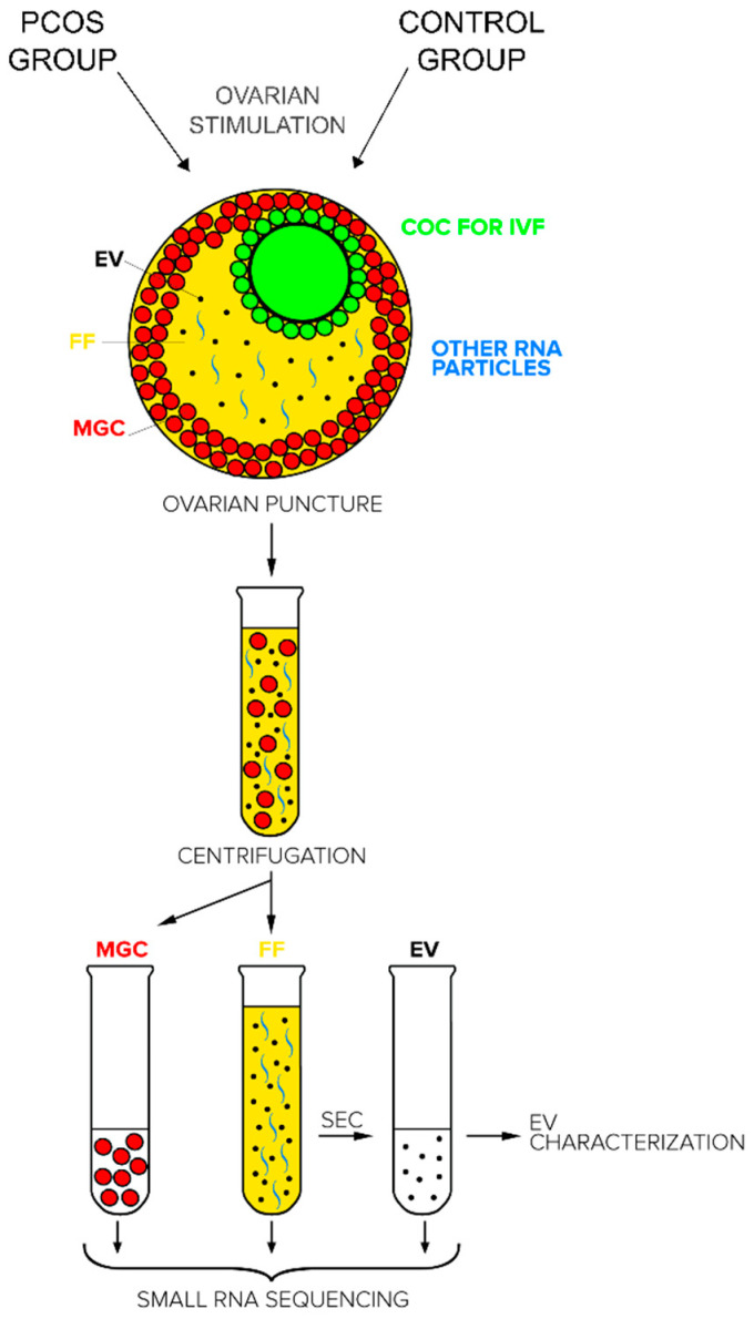
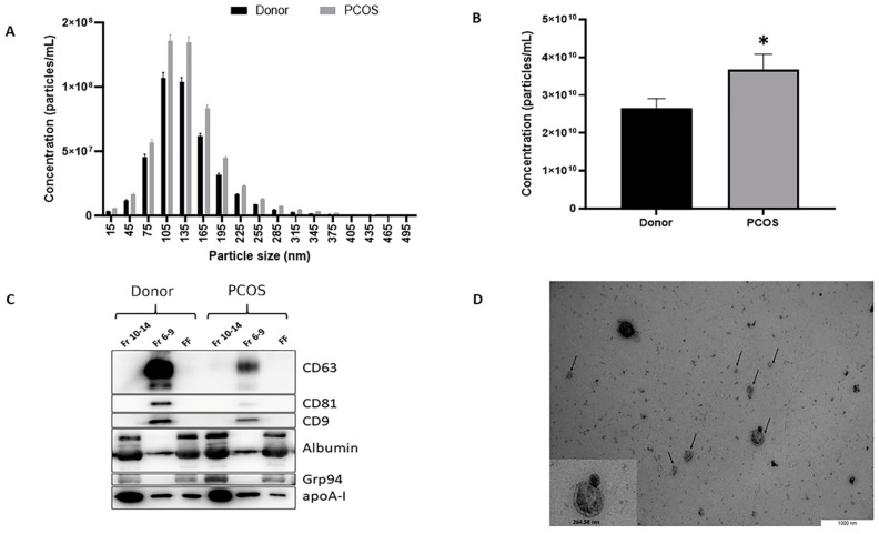
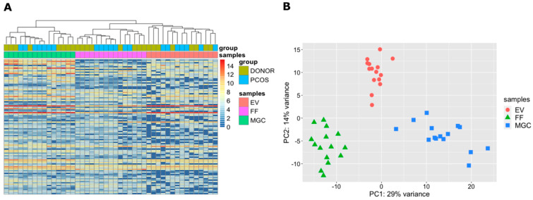
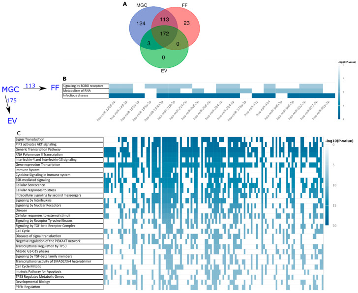
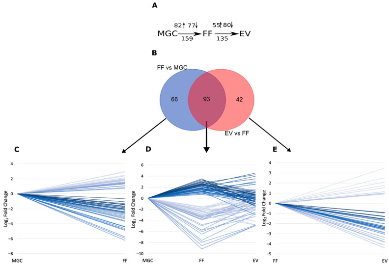
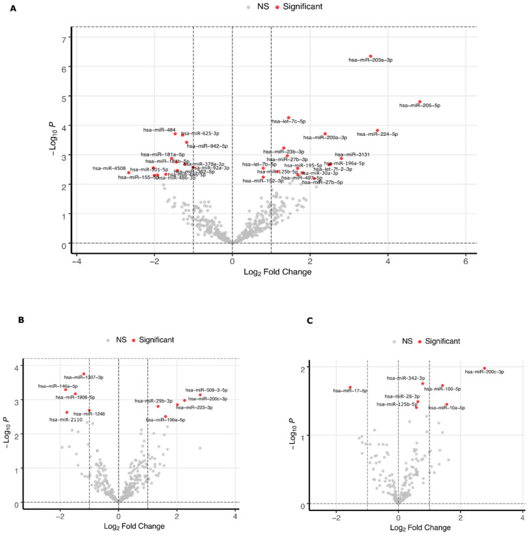
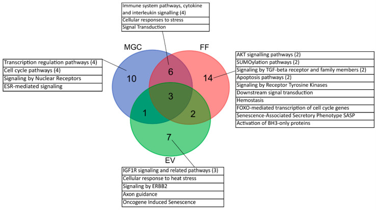
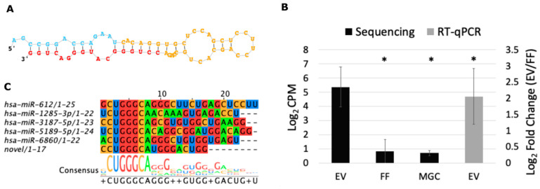
Cell-free RNAs have the potential to act as a means of gene expression regulation between cells and are therefore used as diagnostic markers describing the state of tissue environment. The origin and functions of such RNAs in human ovarian follicle, the environment of oocyte maturation, are unclear. The current study investigates the difference in the microRNA profiles of fertile women and polycystic ovary syndrome (PCOS) patients in three compartments from the same preovulatory follicle: mural granulosa cells (MGC), cell-free follicular fluid (FF), and extracellular vesicles (EV) of the FF by small RNA sequencing. In silico analysis was used for the prediction and over-representation of targeted pathways for the detected microRNAs. PCOS follicles were distinguished from normal tissue by the differential expression of 30 microRNAs in MGC and 10 microRNAs in FF (FDR < 0.1) that commonly regulate cytokine signaling pathways. The concentration of EV-s was higher in the FF of PCOS patients (p = 0.04) containing eight differentially expressed microRNAs (p < 0.05). In addition, we present the microRNA profiles of MGC, FF, and EV in the fertile follicle and demonstrate that microRNAs loaded into EVs target mRNAs of distinct signaling pathways in comparison to microRNAs in FF. To conclude, the three follicular compartments play distinct roles in the signaling disturbances associated with PCOS.

Keywords: PCOS; extracellular vesicles; follicular fluid; granulosa cells; human ovarian follicle; intercellular communication; miRNA; polycystic ovary syndrome.


Conflict of interest statement

The authors declare that the research was conducted in the absence of any commercial or financial relationships that could be construed as a potential conflict of interest.

Figures

Figure 1
Figure 1
Schematic description of sample collection and processing from human pre-ovulatory follicles of polycystic ovary syndrome (PCOS) patients and fertile control group. COC—cumulus oocyte complex (green), FF—cell-free follicular fluid (yellow) containing extracellular vesicles (EV, black) and non-EV-bound RNA (blue), IVF—in vitro fertilization, MGC—mural granulosa cells (red).
Figure 2
Figure 2
Characterization of extracellular vesicles (EVs) isolated from cell-free follicular fluid (FF). (A) Size profile of EVs in study groups (mean ± SEM). (B) Concentration of EVs in polycystic ovarian syndrome patients (PCOS) and control samples (mean ±SEM). (C) Positive signals of EV markers CD63, CD81, CD9 were detected from EV samples (Fr 6-9), while undetectable from protein fractions (Fr 10-14) and FF samples before EV isolation (FF). Albumin, Grp94, and apoA-I were used as markers of negative selection demonstrating diminished signal intensity in EV samples compared to the protein fraction and FF samples. (D) Transmission electron microscopy analysis of purified EVs, indicated by arrows. Data in (A,B) is demonstrated as mean ± SEM. * p = 0.04 Student’s t-test.
Figure 3
Figure 3
Cluster analysis of investigated samples and study groups. (A) Hierarchical clustering of top 100 most variable miRNAs across all samples. Results are depicted by DESeq2 normalized counts on log2 scale. (B) Principal component analysis based on expressed miRNAs per sample type. EV—extracellular vesicles, FF—cell-free follicular fluid, MGC—mural granulosa cells, PCOS—polycystic ovary syndrome.
Figure 4
Figure 4
Cellular and extracellular miRNAs observed in ovarian follicles of fertile women. (A) Distribution of observed miRNAs (>5 reads in >50% of samples) between sample types. (B) Pathways over-represented by the targets of 113 miRNAs secreted by mural granulosa cells (MGC) into follicular fluid (FF) outside of extracellular vesicles (EV). (C) Top 30 pathways potentially regulated by 175 miRNAs secreted into FF in EVs. Each column in (B,C) corresponds to one miRNA regulating a pathway, if marked in blue.
Figure 5
Figure 5
Comparison of cellular and cell-free miRNA expression levels. (A) Schematic representation of miRNA expression level comparisons performed between sample types and the number of differentially expressed (DE) results obtained. Arrows depict the number of upregulated (↑) or downregulated (↓) genes in each comparison. (B) Number of distinct and shared DE miRNAs between comparisons. (C) DE results unique to FF vs. MGC comparisons (D) DE results common for FF vs. MGC and EV vs. FF comparisons. (E) DE results unique to EV vs. FF comparisons. Each line in panels (CE) depicts the statistically significant (FDR < 0.05) expression level difference between sample types of one miRNA. EV—extracellular vesicles, FF—cell-free follicular fluid, MGC – mural granulosa cells.
Figure 6
Figure 6
Differentially expressed miRNAs between oocyte donors and PCOS patients in mural granulosa cell samples (A), cell-free follicular fluid samples (B) and extracellular vesicle samples (C). Statistical significance cut-off is FDR < 0.1 (A and B) or p < 0.05 (C).
Figure 7
Figure 7
Number of Reactome pathways over-represented for miRNAs that are more abundantly expressed in PCOS patients compared to the fertile control group in each sample type. Numbers in brackets refer to combined pathways with similar outcome. EV—extracellular vesicles, FF—cell-free follicular fluid, MGC—mural granulosa cells.
Figure 8
Figure 8
Novel miRNA detected by small RNA sequencing of single follicle components. (A) Predicted stem-loop sequence by miRDeep2. (B) Novel miRNA expression levels in the extracellular vesicles of follicular fluid (EV), cell-free follicular fluid (FF) and in granulosa cells (MGC) according to RNA sequencing displayed as a mean of count per million (CPM) ±SEM on log2 scale (n = 15). RT-qPCR validation result is displayed as fold change ±SEM on log2 scale (n = 15). (C) Novel miRNA aligned to previously annotated miRNAs with similar seed sequence. * p < 0.05, Student’s t-test.

References

    1. Teede H., Deeks A., Moran L. Polycystic ovary syndrome: A complex condition with psychological, reproductive and metabolic manifestations that impacts on health across the lifespan. BMC Med. 2010;8:41. doi: 10.1186/1741-7015-8-41. - DOI - PMC - PubMed
    1. Ehrmann D.A. Polycystic ovary syndrome. N. Engl. J. Med. 2005;352:1223–1236. doi: 10.1056/NEJMra041536. - DOI - PubMed
    1. Goodman N.F., Cobin R.H., Futterweit W., Glueck J.S., Legro R.S., Carmina E. American association of clinical endocrinologists, american college of endocrinology, and androgen excess and pcos society disease state clinical review: Guide to the best practices in the evaluation and treatment of polycystic ovary syndrome—Part 1. Endocr. Pract. 2015;21:1291–1300. doi: 10.4158/EP15748.DSC. - DOI - PubMed
    1. Teede H.J., Misso M.L., Costello M.F., Dokras A., Laven J., Moran L., Piltonen T., Norman R.J. Recommendations from the international evidence-based guideline for the assessment and management of polycystic ovary syndrome. Fertil. Steril. 2018;110:364–379. doi: 10.1016/j.fertnstert.2018.05.004. - DOI - PMC - PubMed
    1. Hennet M.L., Combelles C.M.H. The antral follicle: A microenvironment for oocyte differentiation. Int. J. Dev. Biol. 2012;56:819–831. doi: 10.1387/ijdb.120133cc. - DOI - PubMed