. 2020 Dec 15;21(24):9558.
doi: 10.3390/ijms21249558.

TGF-β Serum Levels in Diabetic Retinopathy Patients and the Role of Anti-VEGF Therapy

Affiliations

TGF-β Serum Levels in Diabetic Retinopathy Patients and the Role of Anti-VEGF Therapy

Vincenza Bonfiglio et al. Int J Mol Sci. .

Abstract

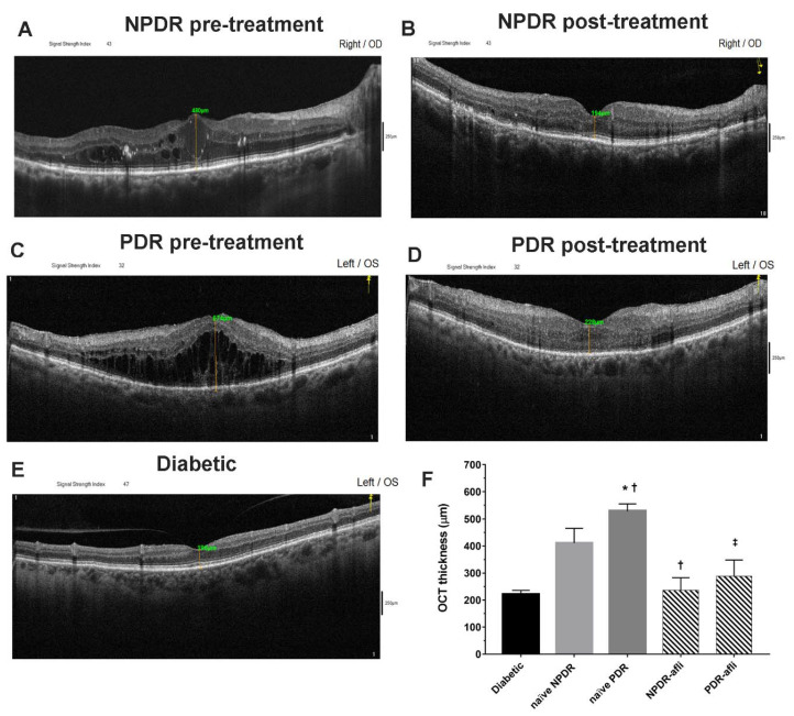
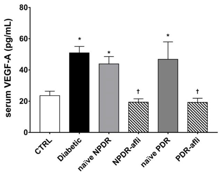
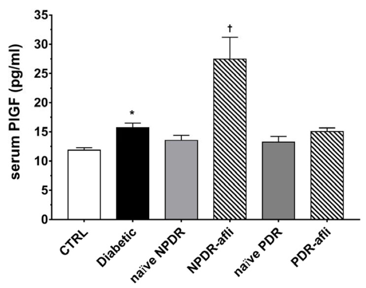
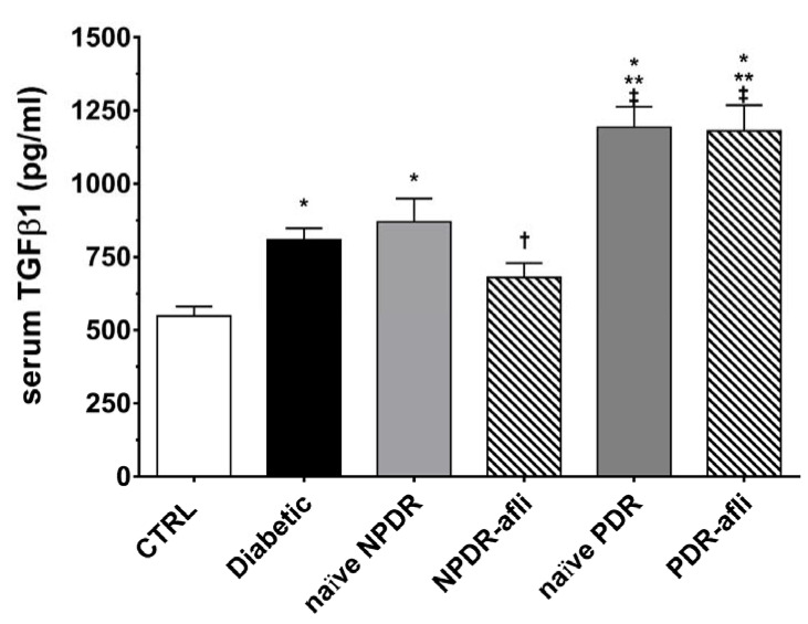
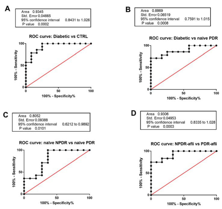
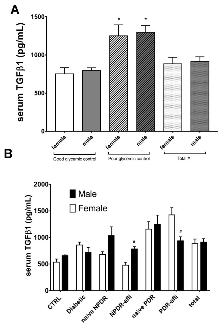
Transforming growth factor β1 (TGFβ1) is a proinflammatory cytokine that has been implicated in the pathogenesis of diabetic retinopathy (DR), particularly in the late phase of disease. The aim of the present study was to validate serum TGFβ1 as a diagnostic and prognostic biomarker of DR stages. Thirty-eight subjects were enrolled and, after diagnosis and evaluation of inclusion and exclusion criteria, were assigned to six groups: (1) healthy age-matched control, (2) diabetic without DR, (3) non-proliferative diabetic retinopathy (NPDR) naïve to treatment, (4) NPDR treated with intravitreal (IVT) aflibercept, (5) proliferative diabetic retinopathy (PDR) naïve to treatment and (6) PDR treated with IVT aflibercept. Serum levels of vascular endothelial growth factor A (VEGF-A), placental growth factor (PlGF) and TGFβ1 were measured by means of enzyme-linked immunosorbent assay (ELISA). Foveal macular thickness (FMT) in enrolled subjects was evaluated by means of structural-optical coherence tomography (S-OCT). VEGF-A serum levels decreased in NPDR and PDR patients treated with aflibercept, compared to naïve DR patients. PlGF serum levels were modulated only in aflibercept-treated NPDR patients. Particularly, TGFβ1 serum levels were predictive of disease progression from NPDR to PDR. A Multivariate ANOVA analysis (M-ANOVA) was also carried out to assess the effects of fixed factors on glycated hemoglobin (HbA1c) levels, TGFβ1, and diabetes duration. In conclusion, our data have strengthened the hypothesis that TGFβ1 would be a biomarker and pharmacological target of diabetic retinopathy.

Keywords: TGFβ; anti-VEGFA; diabetic retinopathy; serum biomarkers.


Conflict of interest statement

Authors have no conflict of interest to declare.

Figures

Figure 1
Figure 1
Representative optical coherence tomography (OCT) images of macular thickness. Aflibercept treatment decreased significantly (p < 0.05) foveal macular thickness in non-proliferative diabetic retinopathy (NPDR) and proliferative diabetic retinopathy (PDR) patients, compared to untreated naïve groups. Foveal macular thickness measurement in enrolled subjects beloging to the followig groups: NPDR before (A) and after (B) aflibercept treatment, PDR before (C) and after (D) aflibercept treatment, and OCT evaluation in diabetic patients without DR (E). Mean foveal macular thickness (F) μm ± S.D.; * p < 0.05 vs. diabetic; † p < 0.05 vs. NPDR naïve; ‡ vs. PDR naïve patients.
Figure 2
Figure 2
VEGF-A serum levels. After 7 days, aflibercept treatment significantly (p < 0.05) decreased VEGFA serum levels in NPDR and PDR patients, compared to diabetic patients without signs of DR, and compared to untreated naïve NPDR and PDR groups. * p < 0.05 vs. CTRL; † naïve vs. aflibercept (afli) treatment.
Figure 3
Figure 3
PlGF serum levels. After 7 days, aflibercept significantly (p < 0.05) increased PlGF serum levels only in NPDR treated patients, compared to other study subject groups. * p < 0.05 vs. CTRL; † naïve vs. aflibercept treatment.
Figure 4
Figure 4
TGFβ1 serum levels. After 7 days, aflibercept significantly (p < 0.05) decreased TGFβ1 serum levels only in NPDR treated patients, compared to other study subject groups. * p < 0.05 vs. CTRL; ** p < 0.05 vs. diabetic patients without signs of DR; † p < 0.05 vs. NPDR naïve patients; ‡ p < 0.05 vs. NPDR either naïve or treated patients.
Figure 5
Figure 5
Receiving-operating characteristics curves for TGFβ1 serum levels. C-statistics validated TGFβ1 serum levels as a predictive biomarker of (A) diabetic patients without sign of DR (diabetic) compared to control healthy subjects; (B) diabetic compared to naïve PDR patients; (C) naïve NPDR compared to naïve PDR patients; (D) NPDR treated with aflibercept (afli) compared to PDR treated (afli) patients.
Figure 6
Figure 6
Glycemic control, group and gender effects on TGFβ1 serum levels. The M-ANOVA post-hoc analysis shed light on fixed factors (independent variables) effects on all analyzed dependent variables. A multivariate-ANOVA guided the stratification of TGFβ1 levels in terms of glycemic control and gender. (A) Both males and females, with reported poor glycemic control at enrollment, showed significantly (p < 0.05) higher levels of TGF1β, compared to other patients. The stratification of TGFβ1 on the basis of gender and subject group (B) showed that only PDR females treated with aflibercept had significantly (p < 0.05) higher levels of TGFβ1, compared to males of the same group. * p < 0.05 vs. “good glycemic control” group; # p < 0.05 males vs. females.

References

    1. Miller D.J., Cascio M.A., Rosca M.G. Diabetic Retinopathy: The Role of Mitochondria in the Neural Retina and Microvascular Disease. Antioxidants. 2020;9:905. doi: 10.3390/antiox9100905. - DOI - PMC - PubMed
    1. Grading Diabetic Retinopathy from Stereoscopic Color Fundus Photographs—An Extension of the Modified Airlie House Classification: ETDRS Report Number 10. Ophthalmology. 2020;98:786–806. doi: 10.1016/j.ophtha.2020.01.030. - DOI - PubMed
    1. Parravano M., De Geronimo D., Scarinci F., Querques L., Virgili G., Simonett J.M., Varano M., Bandello F., Querques G. Diabetic Microaneurysms Internal Reflectivity on Spectral-Domain Optical Coherence Tomography and Optical Coherence Tomography Angiography Detection. Am. J. Ophthalmol. 2017;179:90–96. doi: 10.1016/j.ajo.2017.04.021. - DOI - PubMed
    1. Lee C.S., Lee A.Y., Baughman D., Sim D., Akelere T., Brand C., Crabb D.P., Denniston A.K., Downey L., Fitt A., et al. The United Kingdom Diabetic Retinopathy Electronic Medical Record Users Group: Report 3: Baseline Retinopathy and Clinical Features Predict Progression of Diabetic Retinopathy. Am. J. Ophthalmol. 2017;180:64–71. doi: 10.1016/j.ajo.2017.05.020. - DOI - PMC - PubMed
    1. Bandello F., Cicinelli M.V. 19th EURETINA Congress Keynote Lecture: Diabetic Retinopathy Today. Ophthalmologica. 2020;243:163–171. doi: 10.1159/000506312. - DOI - PubMed

MeSH terms

Substances