. 2021 Feb 19;128(4):492-507.
doi: 10.1161/CIRCRESAHA.120.317348. Epub 2020 Dec 18.

Klotho Deficiency Causes Heart Aging via Impairing the Nrf2-GR Pathway

Affiliations

Klotho Deficiency Causes Heart Aging via Impairing the Nrf2-GR Pathway

Kai Chen et al. Circ Res. .

Erratum in

Abstract

Rationale: Cardiac aging is an important contributing factor for heart failure, which affects a large population but remains poorly understood.

Objective: The purpose of this study is to investigate whether Klotho plays a role in cardiac aging.

Methods and results: Heart function declined in old mice (24 months), as evidenced by decreases in fractional shortening, ejection fraction, and cardiac output. Heart size and weight, cardiomyocyte size, and cardiac fibrosis were increased in old mice, indicating that aging causes cardiac hypertrophy and remodeling. Circulating Klotho levels were dramatically decreased in old mice, which prompted us to investigate whether the Klotho decline may cause heart aging. We found that Klotho gene mutation (KL-/-) largely decreased serum klotho levels and impaired heart function. Interestingly, supplement of exogenous secreted Klotho prevented heart failure, hypertrophy, and remodeling in both old mice and KL (-/-) mice. Secreted Klotho treatment inhibited excessive cardiac oxidative stress, senescence and apoptosis in old mice and KL (-/-) mice. Serum phosphate levels in KL (-/-) mice were kept in the normal range, suggesting that Klotho deficiency-induced heart aging is independent of phosphate metabolism. Mechanistically, Klotho deficiency suppressed GR (glutathione reductase) expression and activity in the heart via inhibition of transcription factor Nrf2 (nuclear factor-erythroid 2 p45-related factor 2). Furthermore, cardiac-specific overexpression of GR prevented excessive oxidative stress, apoptosis, and heart failure in both old and KL (-/-) mice.

Conclusions: Klotho deficiency causes cardiac aging via impairing the Nrf2-GR pathway. Supplement of exogenous secreted Klotho represents a promising therapeutic strategy for aging-associated cardiomyopathy and heart failure.

Keywords: aging; apoptosis; glutathione reductase; heart failure; oxidative stress.


Figures

Figure 1.
Figure 1.. Aging caused heart failure and decreased Klotho levels.
(A) The overall heart size and heart weight-to-body weight ratio. N=7. (B) Cardiomyocytes cross-section area highlighted by WGA staining. N=4. (C) Interstitial fibrosis of the heart highlighted by trichrome staining (blue). N=5. (D) Heart function of the left ventricular measured by MRI. N=7. (E) Klotho expression in the kidney. The target bands were first normalized with GAPDH and then calculated as fold changes vs Adult mice. N=4. (F) Circulating secreted Klotho level. N=6. Data are expressed as mean ± SE and analyzed by nonparametric Mann-Whitney test. Scale bar in panel A is 3 mm, Scale bar in panel B and C lower part is 20 μm, scale bar in panel C upper part is 400 μm. FL-Klotho, full-length transmembrane Klotho; S-Klotho, short-form Klotho (secreted and soluble).
Figure 2.
Figure 2.. Genetic Klotho deficiency impaired heart function and caused cardiac hypertrophy.
Mice were fed on low phosphate diet (LPD). (A) Left ventricular fractional shortening. 3 months WT, N=6. 3 months KL (−/−), N=7. 10 months WT, N=5. 10 months KL (−/−), N=5. (B) Left ventricular ejection fraction. 3 months WT, N=6. 3 months KL (−/−), N=7. 10 months WT, N=5. 10 months KL (−/−), N=5. (C) Left ventricular stroke volume. 3 months WT, N=6. 3 months KL (−/−), N=7. 10 months WT, N=5. 10 months KL (−/−), N=5. (D) Heart rate. 3 months WT, N=6. 3 months KL (−/−), N=7. 10 months WT, N=5. 10 months KL (−/−), N=5. (E) The overall heart size. Scale bar is 3 mm. (F) Heart weight-to-body weight ratio. WT, N=10. KL (−/−), N=6. (G) Left ventricular myocardial mass measured by MRI. 3 months WT, N=6. 3 months KL (−/−), N=7. 10 months WT, N=5. 10 months KL (−/−), N=5. (H) Glutathione reductase expression in the heart. The target band was first normalized with GAPDH and then calculated as fold changes vs WT mice. N=5. (I) Serum phosphate level. WT, N=7. KL (−/−), N=5. Data are expressed as mean ± SE and analyzed by Two-way ANOVA followed by Tukey’s multiple comparisons test for panel A-D and G and nonparametric Mann-Whitney test for panel F, H and I.
Figure 3.
Figure 3.. Secreted Klotho rescued heart aging in KL (−/−) mice.
(A) The overall heart size and weight. N=5. (B) ANP, α-MHC and β-MHC mRNA expression (fetal gene program) measured by real-time RT-PCR. Data were first normalized with GAPDH mRNA and then calculated as fold changes vs the WT mice. N=4. (C) Cardiomyocytes cross-sectional areas highlighted by WGA staining. N=4. (D) Interstitial fibrosis of the heart highlighted by trichrome staining (blue). N=5. (E) Heart function of the left ventricle measured by MRI. N=5. (F) Serum phosphate level. N=5. (G) Cardiac reactive oxygen species highlighted by DHE staining (red). Data were calculated as fold changes vs the WT mice. N=4. (H) Cardiomyocyte apoptosis by TUNEL labeling. TUNEL staining (apoptosis), cTnT staining (cardiomyocyte marker), and the merge of both. N=4. Data are expressed as mean ± SE and analyzed by Two-way ANOVA followed by Tukey’s multiple comparisons test. Scale bar in panel A is 3 mm, Scale bar in panel C, D lower part, G and H is 20 μm, scale bar in panel D upper part is 400 μm.
Figure 4.
Figure 4.. Cardiac-specific overexpression of GR improved heart failure in KL (−/−) mice.
(A) The overall heart size. Scale bar is 3 mm. (B) Heart weight-to-body weight ratio. WT, N=6. KL (−/−), N=8. (C) Left ventricular myocardial mass measured by MRI. WT, N=5. KL (−/−), N=6. (D) Representative images of WGA staining. Scale bar is 20 μm. (E) Cardiomyocytes cross-section areas. N=4. (F) Cardiomyocytes density. N=4. (G) Left ventricular fractional shortening. WT, N=5. KL (−/−), N=6. (H) Left ventricular ejection fraction. WT, N=5. KL (−/−), N=6. (I) Left ventricular stroke volume. WT, N=5. KL (−/−), N=6. (J) Heart rate. WT, N=5. KL (−/−), N=6. Data are expressed as mean ± SE and analyzed by Two-way ANOVA followed by Tukey’s multiple comparisons test.
Figure 5.
Figure 5.. Cardiac-specific overexpression of GR attenuated collagen deposition in the heart of KL (−/−) mice.
(A) Interstitial fibrosis of the heart highlighted by trichrome staining. N=5. (B) Collagen-1 expression in the heart. N=6. (C) Scleraxis homolog A expression in the heart. N=3. The target band was first normalized with GAPDH and then calculated as fold changes vs WT-GFP mice. Data are expressed as mean ± SE and analyzed by Two-way ANOVA followed by Tukey’s multiple comparisons test. Scale bar in panel A lower part is 20 μm, scale bar in panel A upper part is 200 μm.
Figure 6.
Figure 6.. AAV2/9-αMHC-GR treatment increased cardiac GR expression and activity in KL (−/−) mice.
(A) Representative WB of GR. (B) Statistical results of GR expression. The target band was first normalized with GAPDH and then calculated as fold changes vs WT-GFP mice. (C) GSH concentration (D) GSSG concentration. (E) GSH /GSSG ratio. N=6. Data are expressed as mean ± SE and analyzed by Two-way ANOVA followed by Tukey’s multiple comparisons test.
Figure 7.
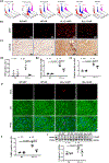
Figure 7.. Cardiac-specific overexpression of GR decreased reactive oxygen species and apoptosis in the heart of KL (−/−) mice.
(A1) Flow cytometry results of ROS. (A2) Statistical results of ROS. N=3. (B1) Representative pictures of DHE staining. (B2) Statistical results of DHE. N=4. (C1) Representative pictures IHC of 4-HNE. (C2) Statistical results of 4-HNE. N=4. (D) Cardiomyocyte apoptosis by TUNEL labeling. TUNEL staining (apoptosis), cTnT staining (cardiomyocyte marker), and the merge of both. (E) Statistical results of TUNEL labelling. N=4. (F) WB results of cleaved-caspase 3 (C-Casp3) expression. The target band was first normalized with GAPDH and then calculated as fold changes vs WT-GFP mice. N=6. Data are expressed as mean ± SE and analyzed by Two-way ANOVA followed by Tukey’s multiple comparisons test. Scale bar is 20 μm.
Figure 8.
Figure 8.. Nrf2 activation is essential for secreted Klotho to regulate glutathione reductase.
(A) Nrf2 and GR protein expression in H9c2 myoblast cells treated with Klotho-free medium (KL (−)) or secreted Klotho (sKL). The target band was first normalized with GAPDH and then calculated as fold changes vs Control (Con). N=4. Data are expressed as mean ± SE and analyzed by Ono-way ANOVA followed by Dunnett’s multiple comparisons test. (B) Nrf2 and GR expression elicited by sKL in the presence or absence of Nrf2 siRNA in H9c2 myoblast cells. The target band was first normalized with GAPDH and then calculated as fold changes vs siRNA Control (siCon). N=3. Data are expressed as mean ± SE and analyzed by Two-way ANOVA followed by Tukey’s multiple comparisons test.

Comment in

References

    1. Hayflick L The future of ageing. Nature. 2000;408:267–9. - PubMed
    1. Hayflick L New approaches to old age. Nature. 2000;403:365. - PubMed
    1. Shih H, Lee B, Lee RJ and Boyle AJ. The aging heart and post-infarction left ventricular remodeling. Journal of the American College of Cardiology. 2011;57:9–17. - PMC - PubMed
    1. Tepp K, Timohhina N, Puurand M, Klepinin A, Chekulayev V, Shevchuk I and Kaambre T. Bioenergetics of the aging heart and skeletal muscles: Modern concepts and controversies. Ageing research reviews. 2016;28:1–14. - PubMed
    1. McMurray JJ and Pfeffer MA. Heart failure. Lancet. 2005;365:1877–89. - PubMed

Publication types

MeSH terms