. 2021 Feb 19;128(4):455-470.
doi: 10.1161/CIRCRESAHA.120.318409. Epub 2020 Dec 18.

JNK2, a Newly-Identified SERCA2 Enhancer, Augments an Arrhythmic [Ca2+]SR Leak-Load Relationship

Affiliations

JNK2, a Newly-Identified SERCA2 Enhancer, Augments an Arrhythmic [Ca2+]SR Leak-Load Relationship

Jiajie Yan et al. Circ Res. .

Abstract

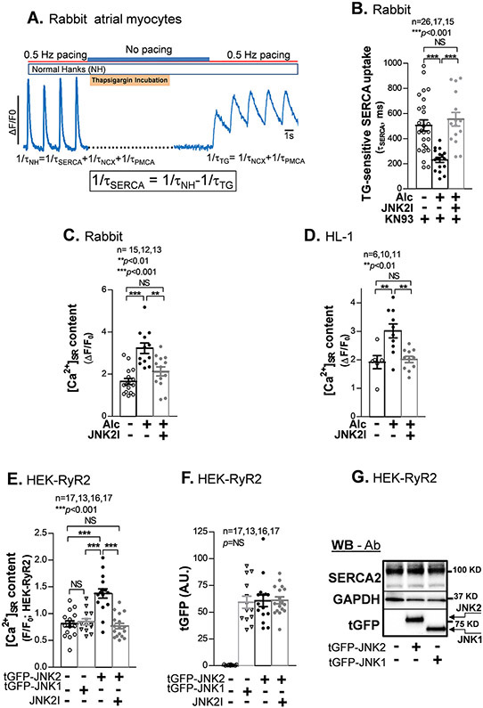
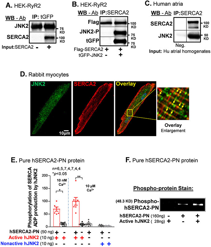
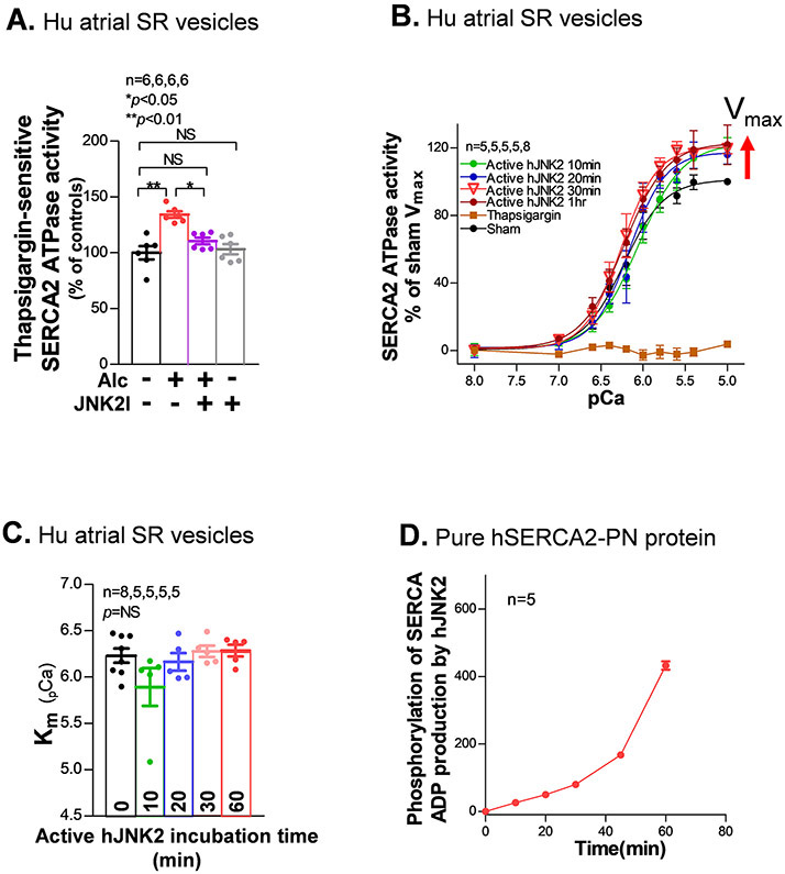
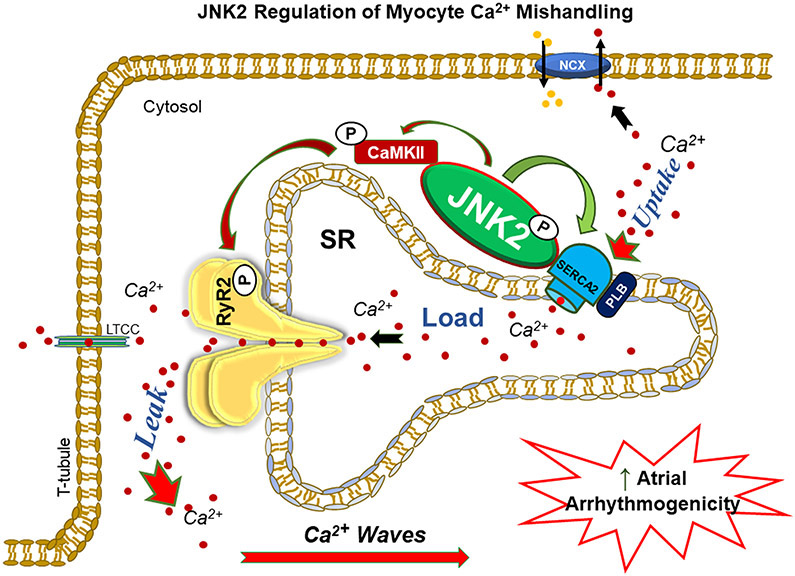
Rationale: We recently discovered pivotal contributions of stress kinase JNK2 (c-Jun N-terminal kinase isoform 2) in increased risk of atrial fibrillation through enhanced diastolic sarcoplasmic reticulum (SR) calcium (Ca2+) leak via RyR2 (ryanodine receptor isoform 2). However, the role of JNK2 in the function of the SERCA2 (SR Ca2+-ATPase), essential in maintaining SR Ca2+ content cycling during each heartbeat, is completely unknown.

Objective: To test the hypothesis that JNK2 increases SERCA2 activity SR Ca2+ content and exacerbates an arrhythmic SR Ca2+ content leak-load relationship.

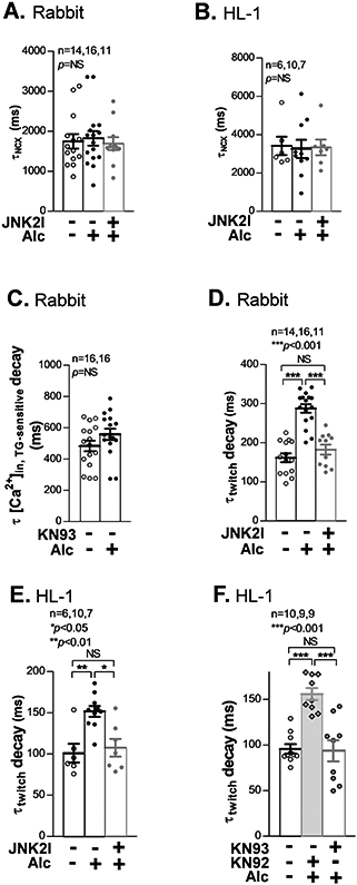
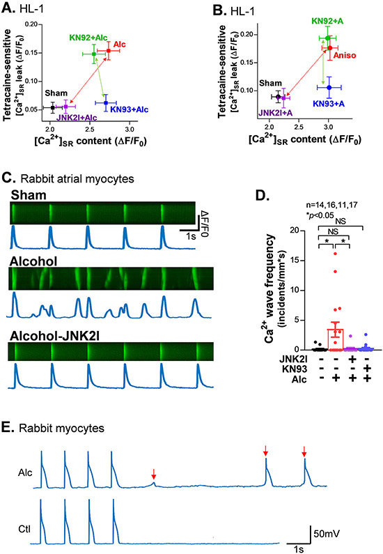
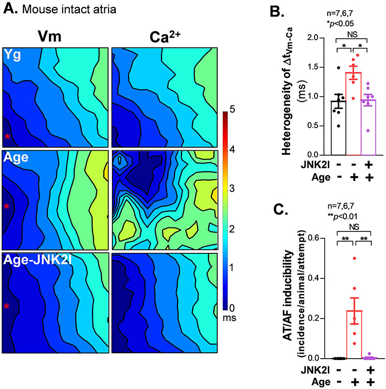
Methods and results: We used confocal Ca2+ imaging in myocytes and HEK-RyR2 (ryanodine receptor isoform 2-expressing human embryonic kidney 293 cells) cells, biochemistry, dual Ca2+/voltage optical mapping in intact hearts from alcohol-exposed or aged mice (where JNK2 is activated). We found that JNK2, but not JNK1 (c-Jun N-terminal kinase isoform 1), increased SERCA2 uptake and consequently elevated SR Ca2+ content load. JNK2 also associates with and phosphorylates SERCA2 proteins. JNK2 causally enhances SERCA2-ATPase activity via increased maximal rate, without altering Ca2+ affinity. Unlike the CaMKII (Ca2+/calmodulin-dependent kinase II)-dependent JNK2 action in SR Ca2+ leak, JNK2-driven SERCA2 function was CaMKII independent (not prevented by CaMKII inhibition). With CaMKII blocked, the JNK2-driven SR Ca2+ loading alone did not significantly raise leak. However, with JNK2-CaMKII-driven SR Ca2+ leak present, the JNK2-enhanced SR Ca2+ uptake limited leak-induced reduction in SR Ca2+, normalizing Ca2+ transient amplitude, but at a higher arrhythmogenic SR Ca2+ leak. JNK2-specific inhibition completely normalized SR Ca2+ handling, attenuated arrhythmic Ca2+ activities, and alleviated atrial fibrillation susceptibility in aged and alcohol-exposed myocytes and intact hearts.

Conclusions: We have identified a novel JNK2-induced activation of SERCA2. The dual action of JNK2 in CaMKII-dependent arrhythmic SR Ca2+ leak and a CaMKII-independent uptake exacerbates atrial arrhythmogenicity, while helping to maintain normal levels of Ca2+ transients and heart function. JNK2 modulation may be a novel therapeutic target for atrial fibrillation prevention and treatment.

Keywords: JNK mitogen-activated protein kinases; animals; atrial fibrillation; phosphorylation; sarcoplasmic reticulum.


Figures

Figure 1.
Figure 1.
JNK2, but not JNK1 increases SR Ca2+ uptake and load. A) An example trace of the thapsigargin (TG)-sensitive SERCA uptake confocal measurement protocol. B) Summarized data showing shortened tau of TG-sensitive Ca2+ removal by SERCA2, reflecting an increased SR Ca2+ uptake, in alcohol (Alc)-exposed rabbit atrial myocytes (in the presence of 1 μM CaMKII inhibitor KN93 to inhibit JNK2-driven diastolic leak) compared to controls. JNK2 inhibition by 170 nM JNK2 inhibitor (JNK2I) reverses the uptake to normal levels seen in controls. C-D) Pooled data of increased [Ca2+]SR load (measured with 10 mM caffeine-induced Ca2+-released transients) in alcohol-exposed rabbit atrial myocytes and HL-1 myocytes; JNK2 specific inhibition by JNK2 inhibitor (JNK2I) completely prevented this alcohol-elevated [Ca2+]SR load. E) Pooled data of a higher [Ca2+]SR load (caffeine-induced Ca2+ transients) in tGFP-JNK2 positively expressed HEK-RyR2 cells (tGFP-JNK2) compared to tGFP-JNK1 positive cells (tGFP-JNK1) and sham controls; the JNK2 action in [Ca2+]SR load is completely eliminated by the JNK2 inhibitor (JNK2I) treatment. F) Summarized quantitative data of tGFP fluorescence intensity from those imaged HEK-RyR2 cells with positively expressed tGFP-JNK1 and tGFP-JNK2 cells and no tGFP in non-transfected controls (Sham). All quantified tGFP-JNK1 and tGFP-JNK2 fluorescence signals were subtracted to non-specific fluorescence background in non-transfected sham control cell images. G) Representative immunoblotting images (reflecting the mean values of quantitative data) showing a similar expression amount of endogenous SERCA2 and exogenous JNK2 and JNK1 proteins (detected by the tGFP antibody) in tGFP-JNK1 and tGFP-JNK2 transfected HEK-RyR cells vs non-transfected controls (Sham).
Figure 2.
Figure 2.
JNK2 is physically associated with SERCA2 and phosphorylates SERCA2. A) Immunoblotting images of co-immunoprecipitated SERCA2 proteins are associated with pull-down tGFP-JNK2 proteins with a tGFP-specific antibody. B) Immunoblotting images of co-immunoprecipitated JNK2 including phosphorylated JNK2 (JNK-P, detected by phospho-specific JNK antibody) and tGFP-tagged JNK2 (detected by tGFP antibody) with SERCA2-specific antibody pull-down Flag-tagged SERCA2 proteins (Flag-SERCA2, detected by Flag antibody) in HEK cells co-transfected with tGFP-JNK2 and Flag-SERCA2 vectors, while negative control (Flag-SERCA2 alone transfection) is in the left lane. C) Immunoblotting images of co-immunoprecipitated SERCA2 proteins with a SERCA2-specific antibody are associated with pull-down JNK2 proteins in human atrial homogenates, while negative control (no SERCA2 antibody) is in the left lane. D) Double immunofluorescence staining of JNK2 and SERCA2 antibodies in rabbit myocytes showing a typical striation pattern of SR for both JNK2 (green) and SERCA2 (red). Far right panel shows an enlarged image of well-colocalized JNK2 and SERCA2 signals (yellow arrows) distributing in a striation pattern. E) Summarized data of increased ADP production of pure active-hJNK2 (but not nonactive-hJNK2) phosphorylation of hSERCA2-PN proteins, with either 10 nM Ca2+ (left panel) or 10 μM Ca2+ (right panel), while the level of ADP production was minimal in the baseline controls of either hJNK2 alone and SERCA alone. F) A Pro-Q Diamond staining gel image of phosphorylated SERCA2-PN proteins (160 ng) by active-hJNK2 (far-right lane) and baseline phosphor-signals of SERCA2-PN alone (middle lane), and undetectable signal of active-hJNK2 alone (far-left lane).
Figure 3.
Figure 3.
JNK2 increases the maximal rate of SERCA2-ATPases activity. A) Summarized data showing alcohol exposure (0.23%, 24hr) increases thapsigargin (TG)-sensitive SERCA2-ATPase activity in isolated human atrial SR vesicles, but the treatment of JNK2-specific inhibitor (JNK2I) attenuates the alcohol-enhanced SERCA2-ATPase activity. JNK2I (170 nM) treatment shows no inhibitory effect on the baseline activity of SERCA2 in sham human atrial SR vesicles. B-C) pooled data of Ca2+-dependent curves of SERCA2-ATPase activity (B) and Ca2+ affinity (Km, C) in human atrial SR vesicles incubated with pure active hJNK2 (64ng) at reaction times of 10 (green), 20 (blue), 30 (pink), and 60 (red) min compared to sham controls (black). And summarized data showing suppressed Ca2+-dependent responses of SERCA2-ATPase activities by incubating with 0.5 μM thapsigargin (brown open squares) compared to sham controls (black dots). E) Pooled data of time-dependent phosphorylation of hSERCA2-PN by 10 ng pure active hJNK2 (10 μM Ca2+).
Figure 4.
Figure 4.
JNK2 increases SERCA2-ATPase activity in a CaMKII-independent manner. A) Summarized data showing active purified human JNK2 (hJNK2) proteins increase thapsigargin-sensitive SERCA2-ATPase activity in isolated human SR vesicles, but additional treatment of CaMKII inhibitor KN93 (1 μM) has no inhibitory effect on this JNK2-enhanced SERCA2-ATPase activity. B) Pooled data of elevated [Ca2+]SR load in 0.23% alcohol (Alc)-exposed (24 hr) HL-1 myocytes with the treatment of CaMKII inhibitor KN93 compared to sham controls (sham) and positive controls treated with KN92 (an inactive analogue of KN93), suggesting lack of action of CaMKII on alcohol-elevated load. C) Summarized data showing CaMKII inhibition has no effect on 50 nM anisomycin (Aniso)-induced increase of [Ca2+]SR load in KN93+Aniso treated as well as KN92+Aniso treated HL-1 myocytes compared to sham controls (sham). D) Summarized data of [Ca2+]SR load in isolated myocytes from alcohol-exposed PLNKO mice showing an increase in [Ca2+]SR load compared to vehicle-treated PLNKO sham controls and JNK2 inhibitor JNK2I treatment precluded this alcohol-evoked increase of [Ca2+]SR load, while lack of PLB in PLNKO mice (vehicle-treated sham controls) markedly increases [Ca2+]SR load compared to WT littermate controls. E) Pooled data of Ca2+-dependent curves of SERCA2-ATPase activity in anti-PLB antibody 2D12 (6 ug) pre-treated human atrial SR vesicles incubated with (pink) and without (blue) pure active hJNK2 (64 ng) compared to sham controls (black).
Figure 5.
Figure 5.
JNK2 prolongs twitch [Ca2+]in decline due to increased diastolic [Ca2+]SR leak (CaMKII-dependent). A-B) Summarized data showing unchanged tau of [Ca2+]in decay of caffeine-induced Ca2+ transients (τNCX), reflecting unchanged contribution of NCX to the diastolic [Ca2+]in removal, in alcohol-exposed rabbit (A) and HL-1 (B) atrial myocytes vs sham controls. C) Summarized data of increased tau of thapsigargin-sensitive [Ca2+]in[Ca]in.TG-sensitive) decay in the absence of KN93, indicating that the presence of CaMKII-dependent [Ca2+]SR leak masks the contribution of [Ca2+]in removal by SERCA2 in 0.23% alcohol-exposed rabbit atrial myocytes compared to controls. D-E) Pooled data of a prolonged tau of [Ca2+]intwitch) decay in alcohol-exposed rabbit myocytes (D) and HL-1 myocytes (E) compared to sham controls; and prevented by 170 nM JNK2I treatment (far right bars). F) Pooled data showing a completely suppressed tau of [Ca2+]intwitch) decay, to the level seen in sham controls (sham), by CaMKII inhibition (1 μM KN93 treatment) in 0.23% alcohol-exposed HL-1 myocytes compared to a significantly prolonged [Ca2+]in decline in alcohol-exposed myocytes treated with 1 μM KN92 (inactive analogue of KN93).
Figure 6.
Figure 6.
JNK2 regulates a [Ca2+]SR load (CaMKII-independent) and leak (CaMKII-dependent) relationship, which driving arrhythmic activities in alcohol-exposed rabbit atrial myocytes. A-B) Plot of the amount of diastolic [Ca2+]SR leak and load in alcohol-exposed (A) and JNK activator anisomycin (Aniso or A)-treated (B) HL-1 myocytes in the presence of 1 μM KN93 or 1 μM KN92 compared to sham controls. Red arrows indicate completely attenuated higher SR Ca2+ leak and load by JNK2-specific inhibition (JNK2I) in both alcohol-exposed and anisomycin (Aniso or A)-treated (B) HL-1 myocytes and green arrows indicate abolished leak but unaffected load by CaMKII inhibition by KN93 but not KN92, an inactive analogue of KN93. C) Representative confocal images of normal Ca2+ transients (0.5Hz) from a sham-control myocyte (upper), multiple Ca2+ waves from an alcohol-exposed myocyte (middle), and normal Ca2+ transients from a JNK2I treated alcohol-exposed atrial myocyte (lower). D) Summarized data showing significantly increased frequency of pacing (0.5Hz)-induced Ca2+ waves in alcohol (Alc)-exposed atrial myocytes compared to sham controls, while these alcohol-evoked changes are eliminated by either JNK2 inhibitor (JNK2I) or CaMKII inhibitor KN93 treatment (far right bars). E) Representative action potential traces of alcohol-exposed (0.23%, 24 hr) and sham control rabbit myocytes showing delayed afterdepolarization (DAD) events at 1Hz electrical pacing, while no DAD events were induced in sham controls. DADs are indicated by the arrows in red.
Figure 7.
Figure 7.
JNK2 drives the heterogeneous relationship between Vm and Ca2+ in aged intact atria. A) Representative isochronal optical maps of simultaneously recorded membrane voltage potentials (Vm; Rh237) and calcium transients (Ca2+; Rhod2) in aged and young atria subjected to a pacing cycle length (CL) of 100 ms (10 Hz). B-C) Summarized data showing a markedly enhanced heterogeneous relationship between Vm and Ca2+ signals. (B) along with increased AF inducibility (C) in aged mouse atria, evidenced by the significantly increased standard deviation of the ΔCa-Vm values from the mapping field vs young controls. And 10 mg/kg JNK2-specific inhibitor (JNK2I) in vivo I.P. treatment in aged WT mice attenuated heterogeneity of ΔCa-Vm and pacing induced AF events (far right bars) compared to untreated aged and young control WT mice.
Figure 8.
Figure 8.
Schematic diagram of demonstrated mechanism of JNK2-driven diastolic leak-uptake relationship, which enhances triggered arrhythmic activities and AF risk.

Comment in

References

    1. Yan J, Zhao W, Thomson JK, Gao X, DeMarco DM, Carrillo E, Chen B, Wu X, Ginsburg KS, Bakhos M, Bers DM, Anderson ME, Song LS, Fill M and Ai X. Stress Signaling JNK2 Crosstalk With CaMKII Underlies Enhanced Atrial Arrhythmogenesis. Circ Res. 2018;122:821–835. - PMC - PubMed
    1. Yan J, Thomson JK, Zhao W, Gao X, Huang F, Chen B, Liang Q, Song LS, Fill M and Ai X. Role of Stress Kinase JNK in Binge Alcohol-Evoked Atrial Arrhythmia. J Am Coll Cardiol. 2018;71:1459–1470. - PMC - PubMed
    1. Go AS, Hylek EM, Phillips KA, Chang Y, Henault LE, Selby JV and Singer DE. Prevalence of diagnosed atrial fibrillation in adults: national implications for rhythm management and stroke prevention: the AnTicoagulation and Risk Factors in Atrial Fibrillation (ATRIA) Study. JAMA. 2001;285:2370–5. - PubMed
    1. Benjamin EJ, Levy D, Vaziri SM, D'Agostino RB, Belanger AJ and Wolf PA. Independent risk factors for atrial fibrillation in a population-based cohort. The Framingham Heart Study. JAMA. 1994;271:840–4. - PubMed
    1. Tonelo D, Providencia R and Goncalves L. Holiday heart syndrome revisited after 34 years. Arquivos brasileiros de cardiologia. 2013;101:183–9. - PMC - PubMed

Publication types

MeSH terms

Substances