. 2020 Jan-Dec:19:1533033820980077.
doi: 10.1177/1533033820980077.

The Effects of Hydrogen Peroxide and/or Radiation on the Survival of Clinically Relevant Radioresistant Cells

Affiliations

The Effects of Hydrogen Peroxide and/or Radiation on the Survival of Clinically Relevant Radioresistant Cells

Yoshikazu Kuwahara et al. Technol Cancer Res Treat. 2020 Jan-Dec.

Abstract

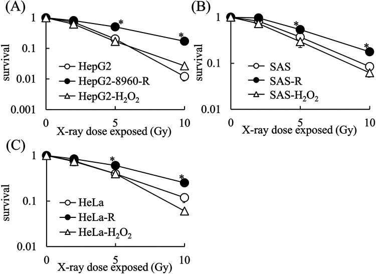
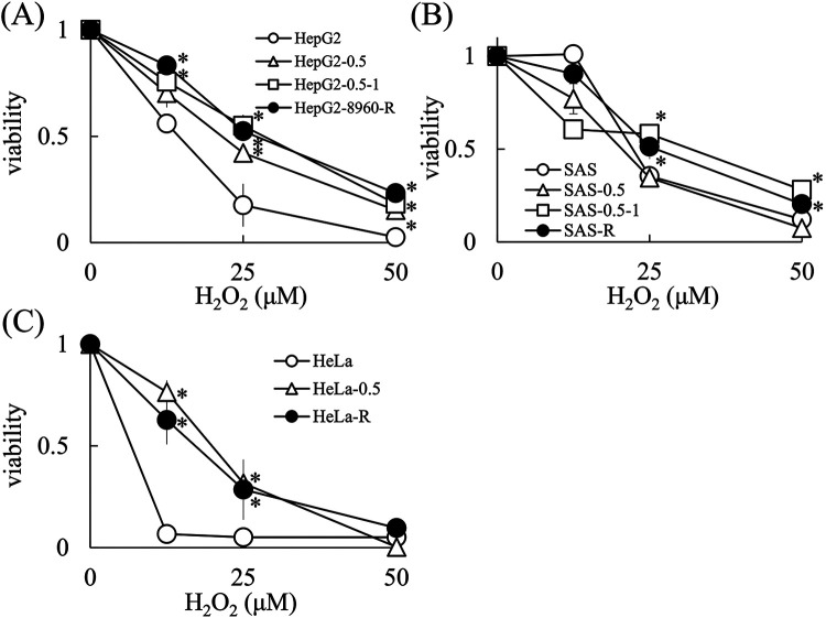
Background: Radiation therapy is a highly cost-effective treatment for cancer, but the existence of radio-resistant cells remains the most critical obstacle in radiotherapy. We have been established clinically relevant radioresistant (CRR) cell lines by exposure to a stepwise increase of fractionated X-rays. We are trying to overcome the radio-resistance by analyzing the properties of these cells. In this study, we tried to evaluate the effects of hydrogen peroxide (H2O2) on the CRR cells because this can evaluate the efficacy of Kochi Oxydol-Radiation Therapy for Unresectable Carcinomas (KORTUC) that treats H2O2 before irradiation. We also established H2O2-resistant cells to compare the radiation and H2O2 resistant phenotype.

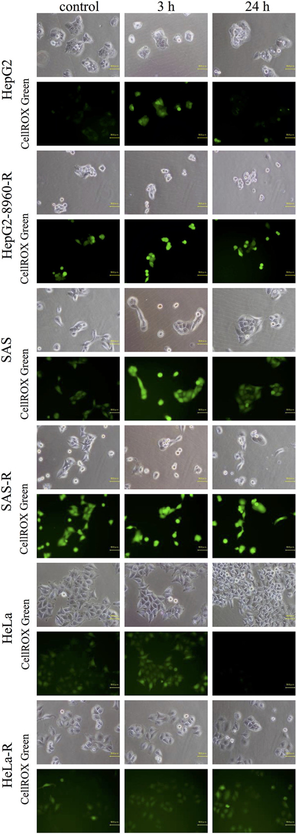
Materials and methods: We used human cancer cell lines derived from hepatoblastoma (HepG2), oral squamous cell carcinoma (SAS), and cervical cancer (HeLa). We established HepG2, SAS, and HeLa CRR cells and HepG2, SAS, and HeLa H2O2-resistant cells. To evaluate their sensitivity to radiation or H2O2, high-density survival assay, or WST assay was performed. CellROXTM was used to detect intracellular Reactive Oxygen Species (ROS).

Results: CRR cells were resistant to H2O2-induced cell death but H2O2-resistant cells were not resistant to irradiation. This phenotype of CRR cells was irreversible. The intracellular ROS was increased in parental cells after H2O2 treatment for 3 h, but in CRR cells, no significant increase was observed.

Conclusion: Fractionated X-ray exposure induces H2O2 resistance in CRR cells. Therefore, it is necessary to carry out cancer therapy such as KORTUC with the presence of these resistant cells in mind, and as the next stage, it would be necessary to investigate the appearance rate of these cells immediately and take countermeasures.

Keywords: H2O2 resistant cells; cell death; clinically relevant radioresistant cells; radiation therapy; reactive oxygen species.


Conflict of interest statement

Declaration of Conflicting Interests: The author(s) declared no potential conflicts of interest with respect to the research, authorship, and/or publication of this article.

Figures

Figure 1.
Figure 1.
CRR cell lines are resistant to H2O2-induced cell death. Cells (5 × 10³) were incubated with H2O2 for 48 hours and a WST assay was performed according to the manufacturer’s protocol. A, H2O2 sensitivity of parental HepG2 and CRR HepG2-8960-R cells. B, H2O2 sensitivity of parental HepG2 and CRR HepG2-R cells. C, H2O2 sensitivity of parental SAS and CRR SAS-R cells. D, H2O2 sensitivity of parental HeLa and CRR HeLa-R cells. The data represent the mean ± SD from 3 independent experiments and are presented relative to the control parental cell lines. *: p < 0.05 using Student’s t test (vs. Parental cells).
Figure 2.
Figure 2.
Generation of H2O2-resistant cell lines. The sensitivity of cells to H2O2-induced cell death was determined by WST assays. The H2O2-resistant cell lines are referred to as their cell line name followed by H2O2. The cells (5 × 10³) were incubated with H2O2 for 48 hours and a WST assay was performed according to the manufacturer’s protocol. A, H2O2 sensitivity of parental HepG2 and HepG2-H2O2 cells. B, H2O2 sensitivity of parental SAS and SAS-H2O2 cells. C, H2O2 sensitivity of parental HeLa and HeLa-H2O2 cells. The data represent the mean ± SD of 3 independent experiments and are presented relative to the control parental cell lines. *: p < 0.05 using Student’s t test (vs. Parental cells).
Figure 3.
Figure 3.
H2O2-resistant cell lines are not resistant to X-ray-induced cell death. A, X-ray sensitivity of HepG2, CRR HepG2-8960-R, and HepG2-H2O2 cells. B, X-ray sensitivity of SAS, CRR SAS-R, and SAS-H2O2 cells. C, X-ray sensitivity of HeLa, CRR HeLa-R, and HeLa-H2O2 cells. The data represent the mean ± SD of 3 independent experiments and are presented relative to the control parental cell lines. *: p < 0.05 using Dunnett’s test (vs. Parental cells).
Figure 4.
Figure 4.
Exposure to fractionated X-rays induced H2O2 resistance in cancer cells. Cells (5 × 10³) were incubated with H2O2 for 48 hours and a WST assay was performed according to the manufacturer’s protocol. A, H2O2 sensitivities of parental HepG2 cell, 0.5 Gy/day × 10 of fractionated X-rays pretreated HepG2 (HepG2-0.5) cell, 0.5 Gy/day × 10 plus 1 Gy/day × 10 of fractionated X-ray pretreated HepG2 (HepG2-0.5-1) cell and CRR HepG2-8960-R cell. B, H2O2 sensitivities of parental SAS, 0.5 Gy/day × 10 of fractionated X-rays pretreated SAS (SAS-0.5) cell, 0.5 Gy/day × 10 plus 1 Gy/day × 10 of fractionated X-ray pretreated SAS (SAS-0.5-1) cell and CRR SAS-R cells. C, H2O2 sensitivity of parental HeLa, 0.5 Gy/day × 10 of fractionated X-rays pretreated HeLa (HeLa-0.5) cell and CRR HeLa-R cell. The data represent the mean ± SD of 3 independent experiments and are presented relative to the control parental cell lines. *: p < 0.05 using Dunnett’s test (vs. Parental cells).
Figure 5.
Figure 5.
The H2O2 resistance phenotype of CRR cell lines was irreversible. Cells (5 × 10³) were incubated with H2O2 for 48 hours and a WST assay was performed according to the manufacturer’s protocol. A, H2O2 sensitivity of parental HepG2, CRR HepG2-8960-R, and HepG2-8960-R-NoIR cells. B, H2O2 sensitivity of parental HepG2, CRR HepG2-R, and HepG2-R-NoIR cells. C, H2O2 sensitivity of parental SAS, CRR SAS-R, and SAS-R-NoIR cells. D, H2O2 sensitivity of parental HeLa, CRR HeLa-R, and HeLa-R-NoIR cells. The data represent the mean ± SD of 3 independent experiments and are presented relative to the control parental cell lines. *: p < 0.05 using Dunnett’s test (vs. Parental cells).
Figure 6.
Figure 6.
Exposure of CRR cells to X-rays enhanced their resistance to H2O2-mediated cell death. Following incubation with H2O2 for 1-h, cells (5 × 10³) was irradiated with 10 Gy of X-rays and incubated extra 48 hours. WST assays were then performed according to the manufacturer’s protocol. A, H2O2 sensitivity of parental HepG2 and HepG2 irradiated cells. B, H2O2 sensitivity of CRR HepG2-8960-R and CRR HepG2-8960-R irradiated cells. C, H2O2 sensitivity of HepG2-R and HepG2-R irradiated cells. D, H2O2 sensitivity of Parental SAS and SAS irradiated cells. E, H2O2 sensitivity of CRR SAS-R and SAS-R irradiated cells. F, H2O2 sensitivity of parental HeLa and HeLa irradiated cells. G, H2O2 sensitivity of CRR HeLa-R and HeLa-R irradiated cells. The data represent the mean ± SD of 3 independent experiments and are presented relative to the control parental or CRR cell lines. *: p < 0.05 using Student’s t test (vs. no irradiated cells).
Figure 7.
Figure 7.
The kinetics of intracellular ROS production in H2O2-treated parental and CRR cells. Cells were treated with or without 100 μM of H2O2 for 3 or 24 hours and the intracellular ROS levels were determined using the CellROX™ Green Reagent according to the manufacturer’s protocol.

References

    1. Bernier J, Hall EJ, Giaccia A. Radiation oncology: a century of achievements. Nat Rev Cancer. 2004;4(9):737–747. doi:10.1038/nrc1451 - PubMed
    1. Kim BM, Hong Y, Lee S, et al. Therapeutic implications for overcoming radiation resistance in cancer therapy. Int J Mol Sci. 2015;16(11):26880–26913. doi:10.3390/ijms161125991 - PMC - PubMed
    1. Tang L, Wei F, Wu Y, et al. Role of metabolism in cancer cell radioresistance and radiosensitization methods. J Exp Clin Cancer Res. 2018;37(1):87 doi:10.1186/s13046-018-0758-7 - PMC - PubMed
    1. Birben E, Sahiner UM, Sackesen C, Erzurum S, Kalayci O. Oxidative stress and antioxidant defense. World Allergy Organ J. 2012;5(1):9–19. doi:10.1097/WOX.0b013e3182439613 - PMC - PubMed
    1. Prise KM, Davies S, Michael BD. Cell killing and DNA damage in Chinese hamster V79 cells treated with hydrogen peroxide. Int J Radiat Biol. 1989;55(4):583–592. doi:10.1080/09553008914550631 - PubMed

Publication types

MeSH terms