

Evidence for a Third Visual Pathway Specialized for Social Perception
- PMID: 33334693
- PMCID: PMC7811363
- DOI: 10.1016/j.tics.2020.11.006
Evidence for a Third Visual Pathway Specialized for Social Perception
Abstract
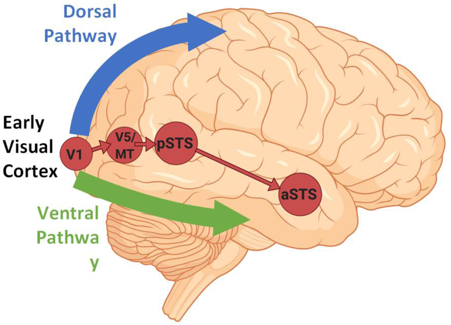
Existing models propose that primate visual cortex is divided into two functionally distinct pathways. The ventral pathway computes the identity of an object; the dorsal pathway computes the location of an object, and the actions related to that object. Despite remaining influential, the two visual pathways model requires revision. Both human and non-human primate studies reveal the existence of a third visual pathway on the lateral brain surface. This third pathway projects from early visual cortex, via motion-selective areas, into the superior temporal sulcus (STS). Studies demonstrating that the STS computes the actions of moving faces and bodies (e.g., expressions, eye-gaze, audio-visual integration, intention, and mood) show that the third visual pathway is specialized for the dynamic aspects of social perception.
Keywords: V5/MT; body perception; face perception; neuroanatomy; social perception; superior temporal sulcus (STS).
Crown Copyright © 2020. Published by Elsevier Ltd. All rights reserved.
Figures

Comment in
-
Developmental Origins of the Pathway for Social Perception.Trends Cogn Sci. 2021 Jul;25(7):546-547. doi: 10.1016/j.tics.2021.03.003. Epub 2021 Mar 16. Trends Cogn Sci. 2021. PMID: 33741276 No abstract available.
-
Third Visual Pathway, Anatomy, and Cognition across Species.Trends Cogn Sci. 2021 Jul;25(7):548-549. doi: 10.1016/j.tics.2021.04.002. Epub 2021 May 20. Trends Cogn Sci. 2021. PMID: 34024730 No abstract available.
Similar articles
-
Characterizing the Third Visual Pathway for Social Perception.Trends Cogn Sci. 2021 Jul;25(7):550-551. doi: 10.1016/j.tics.2021.04.008. Epub 2021 May 20. Trends Cogn Sci. 2021. PMID: 34024729 No abstract available.
-
The Superior Temporal Sulcus Is Causally Connected to the Amygdala: A Combined TBS-fMRI Study.J Neurosci. 2017 Feb 1;37(5):1156-1161. doi: 10.1523/JNEUROSCI.0114-16.2016. Epub 2016 Dec 23. J Neurosci. 2017. PMID: 28011742 Free PMC article.
-
Moving and Static Faces, Bodies, Objects, and Scenes Are Differentially Represented across the Three Visual Pathways.J Cogn Neurosci. 2024 Dec 1;36(12):2639-2651. doi: 10.1162/jocn_a_02139. J Cogn Neurosci. 2024. PMID: 38527070 Free PMC article.
-
One object, two networks? Assessing the relationship between the face and body-selective regions in the primate visual system.Brain Struct Funct. 2022 May;227(4):1423-1438. doi: 10.1007/s00429-021-02420-7. Epub 2021 Nov 18. Brain Struct Funct. 2022. PMID: 34792643 Review.
-
Distributed and interactive brain mechanisms during emotion face perception: evidence from functional neuroimaging.Neuropsychologia. 2007 Jan 7;45(1):174-94. doi: 10.1016/j.neuropsychologia.2006.06.003. Epub 2006 Jul 18. Neuropsychologia. 2007. PMID: 16854439 Review.
Cited by
-
A social cognition perspective on misophonia.Philos Trans R Soc Lond B Biol Sci. 2024 Aug 26;379(1908):20230257. doi: 10.1098/rstb.2023.0257. Epub 2024 Jul 15. Philos Trans R Soc Lond B Biol Sci. 2024. PMID: 39005025 Free PMC article. Review.
-
Looming Angry Faces: Preliminary Evidence of Differential Electrophysiological Dynamics for Filtered Stimuli via Low and High Spatial Frequencies.Brain Sci. 2024 Jan 19;14(1):98. doi: 10.3390/brainsci14010098. Brain Sci. 2024. PMID: 38275518 Free PMC article.
-
The Role of Agentive and Physical Forces in the Neural Representation of Motion Events.J Neurosci. 2024 Jan 10;44(2):e1363232023. doi: 10.1523/JNEUROSCI.1363-23.2023. J Neurosci. 2024. PMID: 38050107 Free PMC article.
-
Neural Correlates Predicting Lane-Keeping and Hazard Detection: An fMRI Study Featuring a Pedestrian-Rich Simulator Environment.Front Hum Neurosci. 2022 Feb 9;16:754379. doi: 10.3389/fnhum.2022.754379. eCollection 2022. Front Hum Neurosci. 2022. PMID: 35221953 Free PMC article.
-
Rethinking simultaneous suppression in visual cortex via compressive spatiotemporal population receptive fields.bioRxiv [Preprint]. 2024 Apr 1:2023.06.24.546388. doi: 10.1101/2023.06.24.546388. bioRxiv. 2024. Update in: Nat Commun. 2024 Aug 11;15(1):6885. doi: 10.1038/s41467-024-51243-7. PMID: 37461470 Free PMC article. Updated. Preprint.
References
-
- Ungerleider LG & Mishkin M 1982. Two cortical visual systems In Ingle DJ, Goodale MA & Mansfield RJW (Eds.) Analysis of Visual Behavior (pp. 549–586). Cambridge, MA: MIT Press.
-
- Milner AD & Goodale MA (1995). The Visual Brain in Action. Oxford: Oxford University Press.
Publication types
MeSH terms
Grants and funding
LinkOut - more resources
Full Text Sources
Other Literature Sources
