Cauda equina compression in metastatic prostate cancer
- PMID: 33334759
- PMCID: PMC7747590
- DOI: 10.1136/bcr-2020-237779
Cauda equina compression in metastatic prostate cancer
Abstract
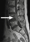
A 67-year-old man presented to his general practitioner with intermittent episodes of unilateral sciatica over a 2-month period for which he was referred for an outpatient MRI of his spine. This evidenced a significant lumbar vertebral mass that showed tight canal stenosis and compression of the cauda equina. The patient was sent to the emergency department for management by orthopaedic surgeons. He was mobilising independently, pain free on arrival and without neurological deficit on assessment. Clinically, this patient presented with no red flag symptoms of cauda equina syndrome or reason to suspect malignancy. In these circumstances, National Institute for Health and Care Excellence guidelines do not support radiological investigation of the spine outside of specialist services. However, in this case, investigation helped deliver urgent care for cancer that otherwise may have been delayed. This leads to the question, do the current guidelines meet clinical requirements?
Keywords: general practice / family medicine; orthopaedic and trauma surgery; prostate; prostate cancer; spinal cord.
© BMJ Publishing Group Limited 2020. No commercial re-use. See rights and permissions. Published by BMJ.
Conflict of interest statement
Competing interests: None declared.
Figures


Similar articles
-
Does a history of lumbar spine surgery predict radiological cauda equina compression in patients undergoing MRI for suspected cauda equina syndrome?Br J Neurosurg. 2020 Feb;34(1):76-79. doi: 10.1080/02688697.2019.1687845. Epub 2019 Nov 13. Br J Neurosurg. 2020. PMID: 31718292
-
Lumbar disc sequestration through the dura into the intrathecal space presenting as acute cauda equina.BMJ Case Rep. 2021 May 5;14(5):e241983. doi: 10.1136/bcr-2021-241983. BMJ Case Rep. 2021. PMID: 33952569 Free PMC article.
-
Evaluation and Management of Cauda Equina Syndrome.Am J Med. 2021 Dec;134(12):1483-1489. doi: 10.1016/j.amjmed.2021.07.021. Epub 2021 Aug 30. Am J Med. 2021. PMID: 34473966 Review.
-
SHADES of grey - The challenge of 'grumbling' cauda equina symptoms in older adults with lumbar spinal stenosis.Musculoskelet Sci Pract. 2020 Feb;45:102049. doi: 10.1016/j.msksp.2019.102049. Epub 2019 Aug 12. Musculoskelet Sci Pract. 2020. PMID: 31439453
-
Cauda equina syndrome.Br J Hosp Med (Lond). 2023 Dec 5;84(11):1-7. doi: 10.12968/hmed.2023.0012. Br J Hosp Med (Lond). 2023. PMID: 38186331 Review.
Cited by
-
Effectiveness and Consequences of Direct Access in Physiotherapy: A Systematic Review.J Clin Med. 2023 Sep 7;12(18):5832. doi: 10.3390/jcm12185832. J Clin Med. 2023. PMID: 37762773 Free PMC article. Review.
References
-
- Koes BW, van Tulder MW, Peul WC. Diagnosis and treatment of sciatica. BMJ 2007;334:1313–7. 10.1136/bmj.39223.428495.BE - DOI - PMC - PubMed
-
- Anthony S. Cauda equina syndrome. Med Prot Soc UK Caseb 2000;20:9–13.
Publication types
MeSH terms
Substances
LinkOut - more resources
Full Text Sources
Medical
Miscellaneous
