

Case Reports
doi: 10.1136/bcr-2020-240193.
Myocardial fibrosis detected by cardiovascular magnetic resonance in absence of myocardial oedema in a patient recovered from COVID-19
Affiliations
- PMID: 33334776
- PMCID: PMC10577794
- DOI: 10.1136/bcr-2020-240193
Item in Clipboard
Case Reports
Myocardial fibrosis detected by cardiovascular magnetic resonance in absence of myocardial oedema in a patient recovered from COVID-19
BMJ Case Rep.
.
No abstract available
Keywords: heart failure; radiology.
Conflict of interest statement
Competing interests: None declared.
Figures
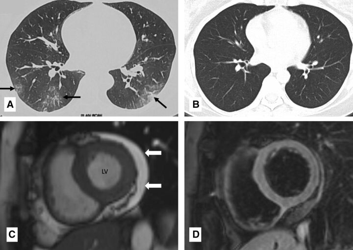
Chest CT at admission and after recovery (A and B) and cardiac magnetic resonance (CMR) at 3 months post recovery from COVID-19 (C and D): axial CT chest at admission (A) showed ground glass opacities (black arrows) in bilateral lower lobes. Axial CT chest 20 days later (B) showed resolution of the lung opacities. CMR short axis cine (balanced steady state free precession) image (C) done 3 months later shows normal left ventricular (LV) size mild pericardial effusion around the LV (white block arrow). CMR fat-saturated T2-weighted image in short axis (D) showed normal signal intensity of the myocardium.
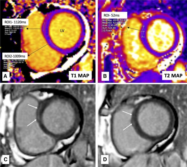
Cardiac magnetic resonance (CMR) tissue characterisation done 3 months after recovery from COVID-19: CMR motion-corrected native T1 mapping image (A) shows raised T1 mapping value (1120 ms) in the basal anteroseptal segment of the left ventricle (LV) (Region of interest, ROI1) and normal value in inferoseptal segment (1009 ms) (ROI2). CMR motion-corrected T2 mapping image (B) shows normal T2 mapping value (52 ms) in the basal anteroseptal segment of LV (with increased T1 values), suggesting the absence of oedema. CMR late gadolinium enhancement (LGE) image in the same section as mapping images (C) and a segment below (D) shows the presence of linear subepicardial LGE (white arrows) in basal anteroseptal segment of the LV, suggestive of replacement fibrosis.