

doi: 10.3174/ajnr.A6904.
Epub 2020 Dec 17.
3D Cranial Nerve Imaging, a Novel MR Neurography Technique Using Black-Blood STIR TSE with a Pseudo Steady-State Sweep and Motion-Sensitized Driven Equilibrium Pulse for the Visualization of the Extraforaminal Cranial Nerve Branches
Affiliations
- PMID: 33334854
- PMCID: PMC7959442
- DOI: 10.3174/ajnr.A6904
Item in Clipboard
3D Cranial Nerve Imaging, a Novel MR Neurography Technique Using Black-Blood STIR TSE with a Pseudo Steady-State Sweep and Motion-Sensitized Driven Equilibrium Pulse for the Visualization of the Extraforaminal Cranial Nerve Branches
AJNR Am J Neuroradiol.
2021 Mar.
Abstract
This study investigated the feasibility of a 3D black-blood STIR TSE sequence with a pseudo steady-state sweep and motion-sensitized driven equilibrium pulse for extraforaminal cranial nerve imaging on a 3T system. Assessments of healthy volunteers showed near-perfect agreement in nerve visualization with excellent to good visualization of the extraforaminal trigeminal, greater occipital, and facial nerves. Suppression of surrounding tissues was excellent to good. 3D cranial nerve imaging can produce nerve selective imaging of extraforaminal cranial and spinal nerve branches.
© 2021 by American Journal of Neuroradiology.
Figures
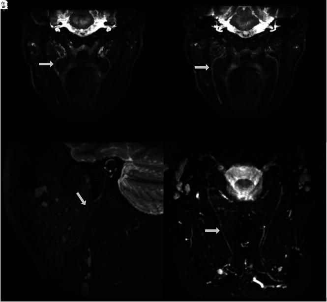
The evaluated cranial and spinal nerve branches acquired using the 3D CRANI sequence. A, Lingual nerve (arrow) on MIP after MPR. B, Inferior alveolar nerve (arrow) after MIP MPR on a coronal oblique reconstruction. C, Extraforaminal facial nerve (arrow) after sagittal oblique MIP MPR, illustrating the intraparotid nerve course. D, Greater occipital nerve (arrow) extending between the semispinalis muscles on MIP MPR.
References
-
- Renton T, Van der Cruyssen F. Diagnosis, pathophysiology, management and future issues of trigeminal surgical nerve injuries. Oral Surg 2019;13:389–403 10.1111/ors.12465 - DOI
MeSH terms
LinkOut - more resources
Full Text Sources
Medical