. 2021 Mar;42(3):573-577.
doi: 10.3174/ajnr.A6911. Epub 2020 Dec 17.

Non-EPI versus Multishot EPI DWI in Cholesteatoma Detection: Correlation with Operative Findings

Affiliations

Non-EPI versus Multishot EPI DWI in Cholesteatoma Detection: Correlation with Operative Findings

J C Benson et al. AJNR Am J Neuroradiol. 2021 Mar.

Abstract

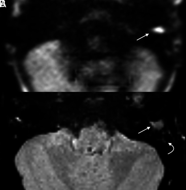
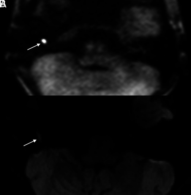
Background and purpose: Although multishot EPI (readout-segmented EPI) has been touted as a robust DWI sequence for cholesteatoma evaluation, its efficacy in disease detection compared with a non-EPI (eg, HASTE) technique is unknown. This study sought to compare the accuracy of readout-segmented EPI with that of HASTE DWI in cholesteatoma detection.

Materials and methods: A retrospective review was completed of consecutive patients who underwent MR imaging for the evaluation of suspected primary or recurrent/residual cholesteatomas. Included patients had MR imaging examinations that included both HASTE and readout-segmented EPI sequences and confirmed cholesteatomas on a subsequent operation. Two neuroradiologist reviewers assessed all images, with discrepancies resolved by consensus. The ratio of signal intensity between the cerebellum and any observed lesion was noted.

Results: Of 23 included patients, 12 (52.2%) were women (average age, 47.8 [SD, 25.2] years). All patients had surgically confirmed cholesteatomas: Six (26.1%) were primary and 17 (73.9%) were recidivistic. HASTE images correctly identified cholesteatomas in 100.0% of patients. On readout-segmented EPI sequences, 16 (69.6%) were positive, 5 (21.7%) were equivocal, and 2 (8.7%) were falsely negative. Excellent interobserver agreement was noted between reviews on both HASTE (κ = 1.0) and readout-segmented EPI (κ = 0.9) sequences. The average signal intensity ratio was significantly higher on HASTE than in readout-segmented EPI, facilitating enhanced detection (mean difference 0.5; 95% CI, 0.3-0.8; P = .003).

Conclusions: HASTE outperforms readout-segmented EPI in the detection of primary cholesteatoma and disease recidivism.


Figures

FIG 1.
FIG 1.
Cholesteatoma with positive findings on both HASTE and RESOLVE in a 4-year-old girl with delayed speech. Both axial HASTE (A) and RESOLVE (B) demonstrate a region of restricted diffusion in the left external auditory canal (arrow). Note greater signal hyperintensity of HASTE compared with the RESOLVE image. Adjacent opacified mastoid air cells demonstrate relatively faint signal (curved arrow).
FIG 2.
FIG 2.
Example of a cholesteatoma seen on HASTE imaging with equivocal findings on RESOLVE. The patient is a 13-year-old boy with a history of bilateral cholesteatomas status post multiple prior surgeries who presented with recurrent hearing loss in his right ear. Axial HASTE (A) image clearly depicts a 0.6-cm focus of restricted diffusion in the right hypotympanum (arrow). Although faint signal is seen in this region on corresponding axial RESOLVE (B) image (arrow), the intralesional signal is insufficiently intense to warrant a certain diagnosis.
FIG 3.
FIG 3.
Cholesteatoma visible on HASTE but not on RESOLVE in a 69-year-old woman who had undergone a left tympanomastoidectomy approximately 15 months prior for resection of a cholesteatoma. MR imaging was performed to assess residual or recurrent disease. Axial (A) HASTE image demonstrates a well-demarcated 0.6-cm focus of restricted diffusion in the left anterior mesotympanum (arrow). No abnormal signal was seen on corresponding RESOLVE image (B).

References

    1. Gilberto N, Custódio S, Colaço T, et al. Middle ear congenital cholesteatoma: systematic review, meta-analysis and insights on its pathogenesis. Eur Arch Otorhinolaryngol Head Neck Dis 2020;277:987–98 10.1007/s00405-020-05792-4 - DOI - PubMed
    1. Nevoux J, Lenoir M, Roger G, et al. Childhood cholesteatoma. Eur Ann Otorhinolaryngol Head Neck Dis 2010;127:143–50 10.1016/j.anorl.2010.07.001 - DOI - PubMed
    1. Olszewska E, Wagner M, Bernal-Sprekelsen M, et al. Etiopathogenesis of cholesteatoma. Eur Arch Otorhinolaryngo l 2004;261:6–24 10.1007/s00405-003-0623-x - DOI - PubMed
    1. Hu Y, Teh BM, Hurtado G, et al. Can endoscopic ear surgery replace microscopic surgery in the treatment of acquired cholesteatoma? A contemporary review. Int J Pediatr Otorhinolaryngol 2020;131:109872 10.1016/j.ijporl.2020.109872 - DOI - PubMed
    1. Choi DL, Gupta MK, Rebello R, et al. Cost-comparison analysis of diffusion weighted magnetic resonance imaging (DWMRI) versus second look surgery for the detection of residual and recurrent cholesteatoma. J Otolaryngol Head Neck Surg 2019;48:58 10.1186/s40463-019-0384-1 - DOI - PMC - PubMed

LinkOut - more resources