

Subcellular Organization of the cAMP Signaling Pathway
- PMID: 33334857
- PMCID: PMC7770493
- DOI: 10.1124/pharmrev.120.000086
Subcellular Organization of the cAMP Signaling Pathway
Abstract
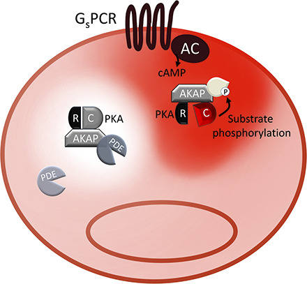
The field of cAMP signaling is witnessing exciting developments with the recognition that cAMP is compartmentalized and that spatial regulation of cAMP is critical for faithful signal coding. This realization has changed our understanding of cAMP signaling from a model in which cAMP connects a receptor at the plasma membrane to an intracellular effector in a linear pathway to a model in which cAMP signals propagate within a complex network of alternative branches and the specific functional outcome strictly depends on local regulation of cAMP levels and on selective activation of a limited number of branches within the network. In this review, we cover some of the early studies and summarize more recent evidence supporting the model of compartmentalized cAMP signaling, and we discuss how this knowledge is starting to provide original mechanistic insight into cell physiology and a novel framework for the identification of disease mechanisms that potentially opens new avenues for therapeutic interventions. SIGNIFICANCE STATEMENT: cAMP mediates the intracellular response to multiple hormones and neurotransmitters. Signal fidelity and accurate coordination of a plethora of different cellular functions is achieved via organization of multiprotein signalosomes and cAMP compartmentalization in subcellular nanodomains. Defining the organization and regulation of subcellular cAMP nanocompartments is necessary if we want to understand the complex functional ramifications of pharmacological treatments that target G protein-coupled receptors and for generating a blueprint that can be used to develop precision medicine interventions.
Copyright © 2020 by The Author(s).
Conflict of interest statement
No author has an actual or perceived conflict of interest with the contents of this article.
Figures
References
-
- Abbattiscianni AC, Favia M, Mancini MT, Cardone RA, Guerra L, Monterisi S, Castellani S, Laselva O, Di Sole F, Conese M, et al. (2016) Correctors of mutant CFTR enhance subcortical cAMP-PKA signaling through modulating ezrin phosphorylation and cytoskeleton organization. J Cell Sci 129:1128–1140. - PubMed
-
- Abrenica B, AlShaaban M, Czubryt MP. (2009) The A-kinase anchor protein AKAP121 is a negative regulator of cardiomyocyte hypertrophy. J Mol Cell Cardiol 46:674–681. - PubMed
-
- Adams SR, Harootunian AT, Buechler YJ, Taylor SS, Tsien RY. (1991) Fluorescence ratio imaging of cyclic AMP in single cells. Nature 349:694–697. - PubMed
Publication types
MeSH terms
Substances
Grants and funding
LinkOut - more resources
Full Text Sources
Other Literature Sources
