Review
. 2020 Dec;8(2):e001408.
doi: 10.1136/jitc-2020-001408.

Phagocytic function of tumor-associated macrophages as a key determinant of tumor progression control: a review

Affiliations
Review

Phagocytic function of tumor-associated macrophages as a key determinant of tumor progression control: a review

Marc Lecoultre et al. J Immunother Cancer. 2020 Dec.

Abstract

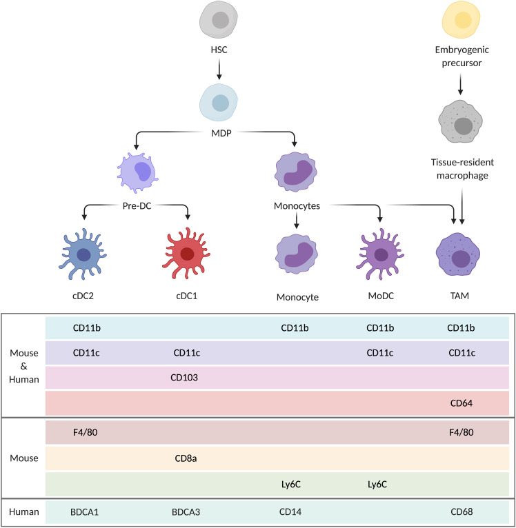
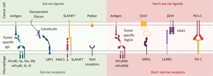
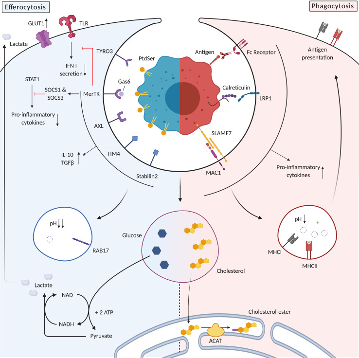
Tumor-associated macrophage (TAM) phagocytic activity is emerging as a new mechanism to harness for cancer treatment. Currently, many approaches are investigated at the preclinical level and some modalities have now reached clinical trials, including the targeting of the phagocytosis inhibitor CD47. The rationale for increasing TAM phagocytic activity is to improve innate anticancer immunity, and to promote T-cell mediated adaptive immune responses. In this context, a clear understanding of the impact of TAM phagocytosis on both innate and adaptive immunity is critical. Indeed, uncertainties persist regarding the capacity of TAM to present tumor antigens to CD8 T cells by cross-presentation. This process is critical for an optimal cytotoxic T-cell immune response and can be mediated by dendritic cells but also potentially by macrophages. In addition, the engulfment of cancer cells affects TAM functionality, as apoptotic cell uptake (a process termed efferocytosis) promotes macrophage anti-inflammatory functions. Because of the abundance of TAM in most solid tumors and the common use of apoptosis inducers such as radiotherapy to treat patients with cancer, efferocytosis potentially affects the overall immune balance within the tumor microenvironment (TME). In this review, we will discuss how cancer cell phagocytosis by TAM impacts antitumor immunity. First, we will focus on the potential of the phagocytic activity of TAM per se to control tumor progression. Second, we will examine the potential of TAM to act as antigen presenting cells for tumor specific CD8 T cells, considering the different characteristics of this process in the tumor tissue and at the molecular level. Finally, we will see how phagocytosis and efferocytosis affect TAM functionality and how these mechanisms impact on antitumor immunity. A better understanding of these aspects will enable us to better predict and interpret the consequences of cancer therapies on the immune status of the TME. Future cancer treatment regimens can thereby be designed to not only impact directly on cancer cells, but also to favorably modulate TAM phagocytic activity to benefit from the potential of this central immune player to achieve more potent therapeutic efficacy.

Keywords: antigen presentation; immunity; innate; macrophages; phagocytosis; tumor microenvironment.


Conflict of interest statement

Competing interests: None declared.

Figures

Figure 1
Figure 1
Mononuclear phagocytes present in tumors and common markers used to distinguish them. HSC give rise to the different subclasses of DC (cDC1, cDC2), monocytes and monocyte-derived DC (MoDC). This common origin causes an overlap regarding the different markers they express. TAM derive from both HSC via monocytes and from tissue-resident macrophages originating from embryogenic precursors. cDC, classical dendritic cells; DC, dendritic cells; HSC, hematopoietic stem cell; MDP, macrophage DC progenitor; MoDC, monocyte-derived DC; TAM, tumor-associated macrophages.
Figure 2
Figure 2
Signals modulating phagocytosis initiation. From left to right: tumor-specific IgG antibodies opsonize cancer cells by binding to tumor antigens. Fcγ receptors (FcγR) expressed on macrophages recognize the constant region of these antibodies and initiate antibody-dependent cell phagocytosis. In humans, the intracellular portion of FcγRI, IIa, IIIa and IIIb possess an immunoreceptor tyrosine-based activation motif (ITAM) that leads to pro-phagocytic activity. In mice, FcγRI, III and IV possess an ITAM. Calreticulin translocation to the surface is induced by cellular stress and DNA damage. Once on the surface of cancer cells, it is stabilized by glycoproteins and glycans and binds to the lipoprotein receptor-related protein 1 (LRP1) on phagocytes. The exact mechanism of action of signaling lymphocytic activation molecule family member 7 (SLAMF7) is unclear, but it promotes cytoskeletal reorganization required for phagocytosis through interaction with macrophage antigen 1 (MAC1) on phagocytes. Phosphatidylserine (PtdSer) is specifically expressed by apoptotic cells and binds to many different receptors among which those of the tumor-associated macrophage (TAM) family (TYRO3, AXL, MerTK) are the best described. It induces efferocytosis, which is a phagocytic process specific for the uptake of apoptotic cells. Eat-me receptor activity is counterbalanced by don’t eat me receptors. First, in both humans and mice, FcγRIIb possesses an immunoreceptor tyrosine-based inhibition motif (ITIM) that negatively regulates initiation of phagocytosis. In humans, IgG4 binds with the highest affinity to FcγRIIb. However, as IgG4 has much greater affinity to the pro-phagocytic FcγRI, the exact role for FcγRIIb expression in vivo is still unknown. In mice, IgG1, IgG2a and IgG2b all bind with low affinity to FcγRIIb. CD47 is the most important “don’t eat me” ligand. SIRPα, its receptor on macrophages, inhibits myosin II polymerization, which is a critical step in initiation of cell engulfment. Major histocompatibility complex class I (MHC I) expression by cancer cells also confers protection against phagocytosis. Leukocyte immunoglobulin-like receptor 1 (LILRB1) binding to the β2-microglobulin component of MHC I prevents phagocytosis. Finally, programmed cell death protein 1 (PD-1) expression on macrophages is correlated with a lower phagocytic activity, which is restored in PD-1 deficient macrophages. Thus, PD-1 activation by its ligand PD-L1 expressed by cancer cells is another inhibitory signal for phagocytosis. Overall, the balance of signaling through eat-me and don’t eat-me receptors will determine initiation of the phagocytic process.
Figure 3
Figure 3
Differences in antigen processing reduce the capacity of macrophages for cross-presentation. Two different cross-presentation pathways have been described: the vacuolar pathway (left) and the cytoplasmic pathway (right). (1) Higher proteolytic activity in the macrophage phagolysosomes leads to a more rapid degradation of ingested antigens and limits their presentation on MHCI. (2) Macrophage capacity to translocate antigens from the phagosome to the cytosol is lower than in DC. As this step is critical for the cytoplasmic pathway, it limits cross-presentation in macrophages. (3) Finally, expression of the cytosolic exonuclease TREX1 is increased in macrophages in response to external stimuli such as irradiation or TLR4 agonists. this limits cGAS-STING pathway activation and type I interferon secretion, which facilitate antigen persistence in phagolysosomes and cross-presentation. cGAS, cyclic GMP-AMP synthase; DC, dendritic cells; ER, endoplasmic reticulum; MHCI, major histocompatibility complex I; STING, stimulator of interferon genes; TAP, transporter associated with antigen processing; TLR, toll-like receptor; TREX1, three-prime repair exonuclease 1.
Figure 4
Figure 4
Efferocytosis promotes anti-inflammatory functions while phagocytosis promotes proinflammatory functions of tumor associated macrophages. Specific receptors for efferocytosis inhibit the downstream signaling of TLR, decrease proinflammatory cytokine secretion through signal transducer and activator of transcription (STAT) 1 inhibition and promote IL-10 and TGFβ secretion. Efferosomes are more acidic than phagosomes, thereby rapidly degrading antigens and limiting the cross-presentation capacity of phagocytes. In contrast, phagocytosis receptors promote an immunogenic response by secreting proinflammatory cytokines and promoting antigen presentation on MHC I and MHC II. Efferocytosis promotes aerobic glycolysis using glucose from engulfed cells but also from the environment through upregulation of the glucose transporter GLUT1. This metabolism contributes to the immunosuppressive tumor microenvironment by depleting glucose and secreting the glycolysis by-product lactate. Potentially cytotoxic quantities of cholesterol are transported to the endoplasmic reticulum where they are esterified by acyl-CoA:cholesterol acyltransferase (ACAT). IFN, interferon; IL, interleukin; LRP1, lipoprotein receptor-related protein 1; NADH, nicotinamide dinucleotide; MAC1, macrophage antigen1; MHC, major histocompatibility complex; SOCS, suppressor of cytokine signaling; SLAMF7, signaling lymphocytic activation molecule family member 7; TGFβ, Transforming growth factor β; TLR, toll-like receptor.

References

    1. Gentles AJ, Newman AM, Liu CL, et al. . The prognostic landscape of genes and infiltrating immune cells across human cancers. Nat Med 2015;21:938–45. 10.1038/nm.3909 - DOI - PMC - PubMed
    1. Cassetta L, Pollard JW. Targeting macrophages: therapeutic approaches in cancer. Nat Rev Drug Discov 2018;17:887–904. 10.1038/nrd.2018.169 - DOI - PubMed
    1. Beltraminelli T, De Palma M. Biology and therapeutic targeting of tumour‐associated macrophages. J Pathol 2020;250:573–92. 10.1002/path.5403 - DOI - PubMed
    1. DeNardo DG, Ruffell B. Macrophages as regulators of tumour immunity and immunotherapy. Nat Rev Immunol 2019;19:369–82. 10.1038/s41577-019-0127-6 - DOI - PMC - PubMed
    1. Mantovani A, Marchesi F, Malesci A, et al. . Tumour-Associated macrophages as treatment targets in oncology. Nat Rev Clin Oncol 2017;14:399–416. 10.1038/nrclinonc.2016.217 - DOI - PMC - PubMed

Publication types