. 2020 Dec 17;11(1):6403.
doi: 10.1038/s41467-020-20125-z.

Metavinculin modulates force transduction in cell adhesion sites

Affiliations

Metavinculin modulates force transduction in cell adhesion sites

Verena Kanoldt et al. Nat Commun. .

Abstract

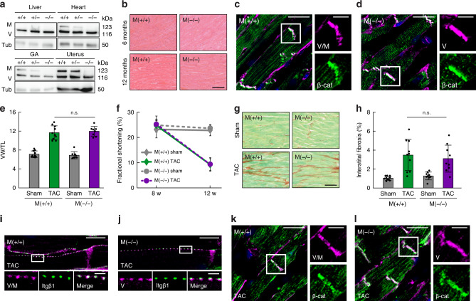
Vinculin is a ubiquitously expressed protein, crucial for the regulation of force transduction in cells. Muscle cells express a vinculin splice-isoform called metavinculin, which has been associated with cardiomyopathies. However, the molecular function of metavinculin has remained unclear and its role for heart muscle disorders undefined. Here, we have employed a set of piconewton-sensitive tension sensors to probe metavinculin mechanics in cells. Our experiments reveal that metavinculin bears higher molecular forces but is less frequently engaged as compared to vinculin, leading to altered force propagation in cell adhesions. In addition, we have generated knockout mice to investigate the consequences of metavinculin loss in vivo. Unexpectedly, these animals display an unaltered tissue response in a cardiac hypertrophy model. Together, the data reveal that the transduction of cell adhesion forces is modulated by expression of metavinculin, yet its role for heart muscle function seems more subtle than previously thought.


Conflict of interest statement

The authors declare no competing interest.

Figures

Fig. 1
Fig. 1. Metavinculin is immobilized in focal adhesions (FAs) and displays enhanced binding to talin.
a Representative images of vinculin-deficient (vinc(−/−)) cells expressing vinculin–venus (V-V) or metavinculin–venus (M-V). Co-staining with a paxillin antibody indicates the independent localization of each vinculin isoform to FAs. b V-V and M-V both rescue the spreading defect of vinc(−/−) cells, which display reduced cellular eccentricity 2 h after cell adhesion when compared to the parental cell line (vinc(f/f)). (n = 23, 32, 27, 30 cells). Two-sided Kolmogorov–Smirnov test: ***p < 0.001. The bar chart shows the mean values ± SD. c V-V and metavinculin–mCherry (M–C) co-localize in FAs when co-expressed in vinc(−/−) cells. d Talin turnover rates are comparable in V-V- or M-V-expressing vinc(−/−) cells, as revealed by FRAP analysis of cells with co-expressed talin-1-TagBFP-HA (T1-B-HA). (n = 12, 12 cells). e Metavinculin resides in FA longer than vinculin as shown by FRAP analysis of vinc(−/−) cells expressing V-V or M-V. (n = 7, 5 cells). f Co-immunoprecipitation (IP) experiments using the venus-tag as bait demonstrate an increased association of talin with metavinculin (IB: immunoblot). g Metavinculin is enriched in an HA-tag-driven IP performed on cells co-expressing T1-B-HA and V-V or M-V. Scale bars indicate 20 µm, in zoom: 5 µm. Expected molecular weight values are indicated (kDa). Source data, exact p values, and uncropped immunoblots with protein markers are provided in the Source Data file.
Fig. 2
Fig. 2. Force transduction in FAs is vinculin isoform-dependent.
a Representative images of vinculin-deficient (vinc(−/−)) cells expressing vinculin tension sensor (V-TS), metavinculin tension sensor (M-TS), and the no-force control (Con-TS) 4 h after spreading on FN-coated glass coverslips show localization of all constructs to FAs (YPet), which are visualized by paxillin staining. Scale bar: 20 µm, in zoom: 5 µm. b Expression of V-TS or M-TS rescues the spreading defect of vinc(−/−) cells; data of the parental (vinc(f/f)) and vinc(−/−) cells are the same as in Fig. 1b. (n = 23, 32, 21, 14 cells). The bar chart shows the mean values ± SD. c Live-cell FLIM measurements of vinc(−/−) cells expressing FL-based tension sensors demonstrate FRET efficiency differences between V-TS and M-TS. Impairing actin binding by inserting the I997A mutation into vinculin (V-TS-I997A) and I1065A into metavinculin (M-TS-I1065A) strongly reduces tension and eliminates vinculin isoform-specific differences. (n = 73, 73, 74, 72, 73 cells). d Live-cell FLIM measurements of FL-based Con-TS, V-TS, and M-TS expressed in talin-deficient cells (tln1−/−tln2−/−), seeded on pLL-coated dishes and treated with Y-27632, confirmed that FRET differences are force-specific. (n = 80, 80, 82 cells). e Highly similar FRET efficiencies of force-insensitive vinculin (V-F7-TS) and metavinculin (M-F7-TS) tension sensor controls expressed in vinc(−/−) cells demonstrate that vinculin isoform-specific effects are conformation-independent (n = 86, 85 cells). f Talin-2 tension sensor (T2-TS) measurements in vinc(−/−) cells expressing TagBFP-tagged vinculin (V-B) and metavinculin (M-B) show that vinculin isoform-specific force transduction propagates across talin-2. T2-Con: talin-2 no-force control. (n = 30, 36, 31, 31 cells). g, h The A50I point mutation, which reduces the binding affinity of (meta)vinculin to talin, caused a FRET efficiency decrease in FL- and F40-based vinculin (V-TS-A50I) and metavinculin (M-TS-A50I) samples. (g: n = 84, 83, 78, 85, 86 cells; h: n = 60, 59, 77, 57, 78 cells). i Examination of stretched sensor molecules in V-TS-expressing cells, using four different TS modules, shows that vinculin is exposed to a wide range of forces; in average, 20–30% of molecules experience mechanical tension (n = 77, 73, 81, 77 cells). j Analogous analysis of M-TS-expressing cells indicates that the fraction of mechanically engaged metavinculin molecules is <20%. Note the equal amounts of stretched molecules in samples containing sensors sensitive to 1–‍6 pN, 3–‍5 pN, and 6–‍8 pN indicating comparably high force per molecule across metavinculin. (n = 80, 74, 80, 77 cells). k Analyzing the differences of medians shown in (i) and (j) indicates that cells expressing M-TS instead of V-TS have less mechanically-engaged linkages that experience higher tension per molecule. Boxplots show median, 25th and 75th percentile with whiskers reaching to the last data point within 1.5× interquartile range. Two-sided Kolmogorov–Smirnov test: ***p < 0.001, **p < 0.01, *p < 0.05, n.s. (not significant) p ≥ 0.05. Source data and exact p values are provided in the Source Data file.
Fig. 3
Fig. 3. Vinculin isoform-specific differences in force transduction are observed in FAs and adherens junctions (AJs) of muscle cells.
a Representative images of HL-1 cells expressing FL-based vinculin tension sensor (V-TS), metavinculin tension sensor (M-TS), and the no-force control (Con-TS) show localization of all fusion proteins to FAs (YPet), which are co-stained with a paxillin antibody. Nuclei are visualized with DAPI (blue). b FLIM measurements of FAs in HL-1 cells expressing FL-based sensors demonstrate vinculin isoform-specific force transduction also in muscle cells. (n = 81, 92, 84 cells). c Representative images of HL-1 cells expressing V-TS, M-TS, and Con-TS show localization of all tension sensors to AJs (YPet). The signal of co-stained α-catenin (α-cat) is used to outline AJs. d FLIM measurements of AJs in HL-1 cells reveal vinculin isoform-specific force transduction in cell–cell contacts. (n = 65, 53, 48 cells). Boxplots show median, 25th, and 75th percentile with whiskers reaching the last data point within 1.5× interquartile range. Two-sided Kolmogorov–Smirnov test: ***p < 0.001, **p < 0.01, *p < 0.05. Scale bar: 20 µm, in zoom: 5 µm. Source data and exact p values are provided in the Source Data file.
Fig. 4
Fig. 4. Metavinculin knockout mice display a normal hypertrophic response upon transverse aortic constriction (TAC).
a Western blot analysis of tissue lysates from 6-month-old mice shows complete loss of metavinculin in knockout M(−/−) and ~50% reduction of expression in heterozygous M(+/−) animals, compared to wild-type M(+/+) littermates. (GA: gastrocnemius muscle, V: vinculin, M: metavinculin, Tub: tubulin). b Histological analysis of M(+/+) and M(−/−) heart tissue of 6- and 13-month-old mice indicates normal morphology of the cardiac muscle. Scale bar: 40 µm. c, d Representative immunohistochemistry of ventricular tissue from 13-month-old M(+/+) and M(−/−) mice, respectively, reveals unchanged intercalated disk (ICD) structures as indicated by β-catenin (β-cat) and (meta)vinculin (V/M) staining. Nuclei are visualized with DAPI (blue). Scale bar: 20 µm, in zoom: 10 µm. e Heart weight analysis of sham- or TAC-operated mice, expressed as the ventricular weight (VW) normalized to tibia length (TL) shows no difference between M(+/+) and M(−/−) mice (n = 10, 10, 9, 9 mice). The bar chart shows the mean values ± SD. f Echocardiography assessment shows the expected decrease in fractional shortening in TAC-operated animals; the effect is highly similar in M(+/+) and M(−/−) mice. (n = 10, 9, 9, 7 mice). The line chart shows mean values ± SD. g Representative images of Sirius Red/Fast Green-stained myocardium show anticipated tissue fibrosis in each group of TAC-operated animals. Scale bar: 40 µm. h Quantitative analysis of interstitial fibrosis reveals a normal tissue response in metavinculin-deficient mice. (n = 10, 10, 9, 9 mice). i, j Representative immunohistochemistry of ventricular tissues from 3-month-old M(+/+) and M(−/−) TAC-operated mice, respectively. Costameres are visualized with β1 integrin (Itgβ1) and (meta)vinculin (V/M) staining. Scale bar: 10 µm, in zoom: 2 µm. k, l Representative immunohistochemistry of ventricular tissues from 3-month-old M(+/+) and M(−/−) TAC-operated mice shows normal ICD structures as indicated by β-catenin (β-cat) and (meta)vinculin (V/M) staining. Nuclei are visualized with DAPI (blue). Scale bar: 20 µm, in zoom: 10 µm. Two-sided ANOVA followed by Sidak’s test: n.s. (not significant) p ≥ 0.05. Expected molecular weight values are indicated (kDa). Source data, exact p values, and uncropped immunoblots with protein markers are provided in the Source Data file.

References

    1. Hoffman BD, Grashoff C, Schwartz MA. Dynamic molecular processes mediate cellular mechanotransduction. Nature. 2011;475:316–323. doi: 10.1038/nature10316. - DOI - PMC - PubMed
    1. Kanoldt V, Fischer L, Grashoff C. Unforgettable force—crosstalk and memory of mechanosensitive structures. Biol. Chem. 2019;400:687–698. doi: 10.1515/hsz-2018-0328. - DOI - PubMed
    1. Geiger B, Spatz JP, Bershadsky AD. Environmental sensing through focal adhesions. Nat. Rev. Mol. Cell Biol. 2009;10:21–33. doi: 10.1038/nrm2593. - DOI - PubMed
    1. Humphries JD, et al. Vinculin controls focal adhesion formation by direct interactions with talin and actin. J. Cell Biol. 2007;179:1043–1057. doi: 10.1083/jcb.200703036. - DOI - PMC - PubMed
    1. Grashoff C, et al. Measuring mechanical tension across vinculin reveals regulation of focal adhesion dynamics. Nature. 2010;466:263–266. doi: 10.1038/nature09198. - DOI - PMC - PubMed

Publication types

MeSH terms