. 2020 Dec 17;10(1):22177.
doi: 10.1038/s41598-020-79324-9.

The phosphoenolpyruvate carboxykinase (PEPCK) inhibitor, 3-mercaptopicolinic acid (3-MPA), induces myogenic differentiation in C2C12 cells

Affiliations

The phosphoenolpyruvate carboxykinase (PEPCK) inhibitor, 3-mercaptopicolinic acid (3-MPA), induces myogenic differentiation in C2C12 cells

Madelaine C Brearley et al. Sci Rep. .

Abstract

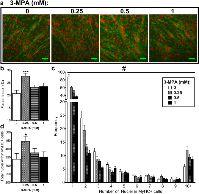
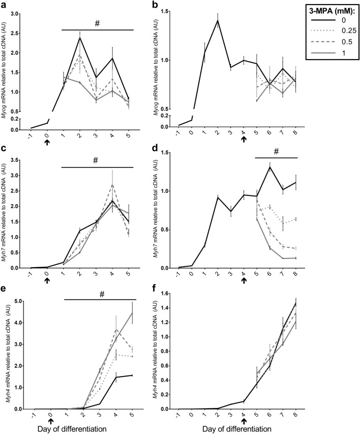
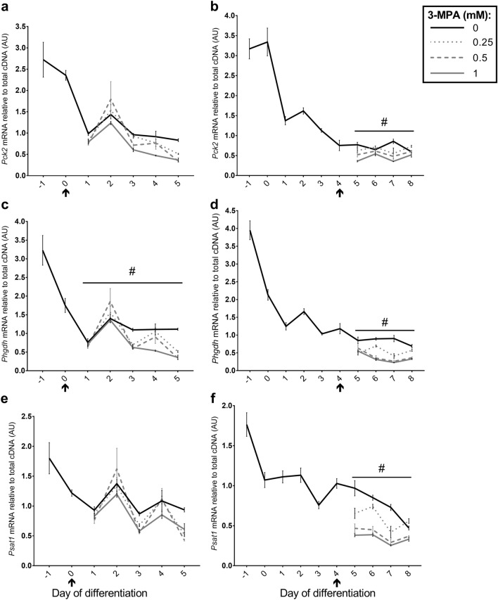
Phosphoenolpyruvate carboxykinase (PEPCK) is a gluconeogenic enzyme with a cytosolic (Pck1/PEPCK-C) and mitochondrial (Pck2/PEPCK-M) isoform. Here we investigate the effect of 3-mercaptopicolinic acid (3-MPA), a PEPCK inhibitor, on C2C12 muscle cells. We report that Pck2 mRNA is 50-5000-fold higher than Pck1 during C2C12 myogenesis, indicating Pck2 is the predominant PEPCK isoform. C2C12 cell proliferation was inhibited in a dose-dependent manner following 48 h 3-MPA treatment (0.01-1 mM). C2C12 myogenic differentiation was significantly induced following 3-MPA treatment (0.25, 0.5, 1 mM) from day 0 of differentiation, demonstrated by increased creatine kinase activity, fusion index and myotube diameter; likewise, the myosin heavy chain (MyHC)-IIB isoform (encoded by Myh4) is an indicator of hypertrophy, and both porcine MYH4-promoter activity and endogenous Myh4 mRNA were also significantly induced. High doses (0.5 and/or 1 mM) of 3-MPA reduced mRNA expression of Pck2 and genes associated with serine biosynthesis (Phosphoglycerate dehydrogenase, Phgdh; phosphoserine aminotransferase-1, Psat1) following treatment from days 0 and 4. To conclude, as Pck2/PEPCK-M is the predominant isoform in C2C12 cells, we postulate that 3-MPA promoted myogenic differentiation through the inhibition of PEPCK-M. However, we were unable to confirm that 3-MPA inhibited PEPCK-M enzyme activity as 3-MPA interfered with the PEPCK enzyme assay, particularly at 0.5 and 1 mM.


Conflict of interest statement

The authors declare no competing interests.

Figures

Figure 1
Figure 1
Treatment with 3-MPA reduced C2C12 cell number by inhibiting proliferation. Proliferating C2C12 cells were treated with a range of 3-MPA doses (0.01–1 mM) or equivalent volume of vehicle control (PBS; 0 mM 3-MPA) diluted in GM supplemented with 5% (v/v) FBS. (a) Cells were collected for total DNA quantification after 0, 8, 24 and 48 h of treatment. Data presented as Means (n = 16) ± SEM. Two-way ANOVA was performed: #P < 0.001 (time × treatment interaction). Cells were also either (b) collected to quantify total DNA content after 48 h or (c) incubated with BrdU labelling solution for the final 3 h of the 48 h period to quantify BrdU incorporation. The data presented in (a) and (b) were obtained in separate experiments. Data presented as Means (n = 8) ± SEM. One-way ANOVA was performed, followed by a Dunnett’s multiple comparison to vehicle control (0 mM 3-MPA): *P < 0.05, **P < 0.01 and ***P < 0.001. 2% and 10% (v/v) FBS were internal controls and therefore excluded from the statistical analyses.
Figure 2
Figure 2
Treatment with 3-MPA induces myogenic differentiation. C2C12 cells were treated with 0, 0.25, 0.5 or 1 mM 3-MPA from day 0 of differentiation for five days, with DM refreshed every 48 h. Samples were collected every 24 h for six days and a Creatine Kinase (CK) activity assay performed. (a) Representative bright-field images captured at day 5 of treatment with 0, 0.25, 0.5 or 1 mM 3-MPA. Photographs were captured at ×6.3 magnification. Scale bar: 100 µm. Data presented as Means (n = 3, except day 0 n = 6) ± SEM for (b) CK activity normalised to DNA content. #P < 0.05 (time × treatment interaction). C2C12 cells were transfected with MYH4-ZsGreen expression plasmid on day − 1, then treated with 0, 0.25 or 1 mM 3-MPA from day 0 until day 4 of differentiation. Measurements were taken for (c) porcine MYH4-promoter activity and (d) average myotube diameter. Data presented as Means ± SEM, from three wells for MYH4-promoter activity (n = 3) and five separate fields of view per well for myotube diameter (n = 15). (c) Student’s t-test was used for dbcAMP treatment and one-way ANOVA for 3-MPA doses and (d) one-way ANOVA (blocking for ‘well’) were performed for each treatment group and appropriate vehicle controls, followed by a post-hoc Dunnett’s multiple comparison test for 3-MPA doses compared to the vehicle control (PBS; 0 mM 3-MPA). *P < 0.05, ***P < 0.001.
Figure 3
Figure 3
Treatment with 3-MPA increases fusion index. C2C12 cells were treated with 0, 0.25, 0.5 or 1 mM 3-MPA from day 0 of differentiation, then after 72 h treatment immunofluorescence was performed using MyHC antibody (green) and PI (red). (a) Photographs captured at 10X magnification. Scale bar: 100 µm. ImageJ software was used to quantify (b) Fusion Index, (c) the frequency of nuclei found within MyHC+ cells and (d) total nuclei number within MyHC  cells. Data presented as Means (n = 12, from four wells per treatment and three fields of view per well) ± SEM. One- or two-way ANOVAs (blocking for ‘well’) were performed as appropriate, followed by Dunnett’s comparison test to the vehicle control (PBS; 0 mM 3-MPA). #P < 0.001 (treatment × nuclei number interaction) *P < 0.05, ***P < 0.001.
Figure 4
Figure 4
Effect of 3-MPA treatment on genes associated with myogenesis. Relative expression of (a, b) Myog (Myogenin), (c, d) Myh7 (encodes Myosin Heavy Chain (MyHC)-I), and (e, f) Myh4 (encodes MyHC-IIB) mRNA following treatment of C2C12 cells with 0, 0.25, 0.5 or 1 mM 3-MPA from day 0 (a, c, e) or day 4 (b, d, f) of differentiation. Data presented as Means (n = 4–6) ± SEM. #P < 0.05 (time × treatment interaction). Start of treatment indicated by ↑. See Supplementary Tables 1 and 2 for Dunnett’s Multiple Comparison tests.
Figure 5
Figure 5
Effect of 3-MPA treatment on genes associated with gluconeogenesis and serine biosynthesis. Relative expression for (a, b) Pck2 (encodes mitochondrial PEPCK isoform), (c, d) phosphoglycerate dehydrogenase (Phgdh) and (e, f) phosphoserine aminotransferase-1 (Psat1) mRNA following treatment of C2C12 cells with 0, 0.25, 0.5 or 1 mM 3-MPA from day 0 (a, c, e) or day 4 (b, d, f) of differentiation. Data presented as Means (n = 4–6) ± SEM. #P < 0.05 (time × treatment interaction). Start of treatment indicated by ↑. See Supplementary Tables 1 and 2 for Dunnett’s Multiple Comparison tests.

References

    1. Brown DM, et al. Mitochondrial phosphoenolpyruvate carboxykinase (PEPCK-M) and serine biosynthetic pathway genes are co-ordinately increased during anabolic agent-induced skeletal muscle growth. Sci. Rep. 2016;6:28693. doi: 10.1038/srep28693. - DOI - PMC - PubMed
    1. Brown D, et al. The Beta-adrenergic agonist, Ractopamine, increases skeletal muscle expression of Asparagine Synthetase as part of an integrated stress response gene program. Sci. Rep. 2018;8:15915. doi: 10.1038/s41598-018-34315-9. - DOI - PMC - PubMed
    1. Brearley MC, et al. Changes in expression of serine biosynthesis and integrated stress response genes during myogenic differentiation of C2C12 cells. Biochem. Biophys. Rep. 2019;20:100694. - PMC - PubMed
    1. Yang J, Kalhan SC, Hanson RW. What is the metabolic role of phosphoenolpyruvate carboxykinase? J. Biol. Chem. 2009;284:27025–27029. doi: 10.1074/jbc.R109.040543. - DOI - PMC - PubMed
    1. Méndez-Lucas A, Hyroššová P, Novellasdemunt L, Viñals F, Perales JC. Mitochondrial phosphoenolpyruvate carboxykinase (PEPCK-M) is a pro-survival, endoplasmic reticulum (ER) stress response gene involved in tumor cell adaptation to nutrient availability. J. Biol. Chem. 2014;289:22090–22102. doi: 10.1074/jbc.M114.566927. - DOI - PMC - PubMed

Publication types

MeSH terms