. 2020 Dec 17;10(1):22211.
doi: 10.1038/s41598-020-79278-y.

Bone marrow stromal cell therapy improves survival after radiation injury but does not restore endogenous hematopoiesis

Affiliations

Bone marrow stromal cell therapy improves survival after radiation injury but does not restore endogenous hematopoiesis

Miguel F Diaz et al. Sci Rep. .

Erratum in

Abstract

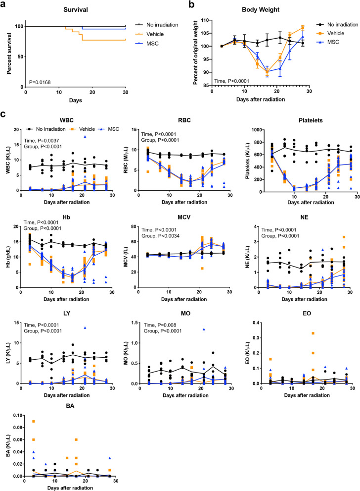
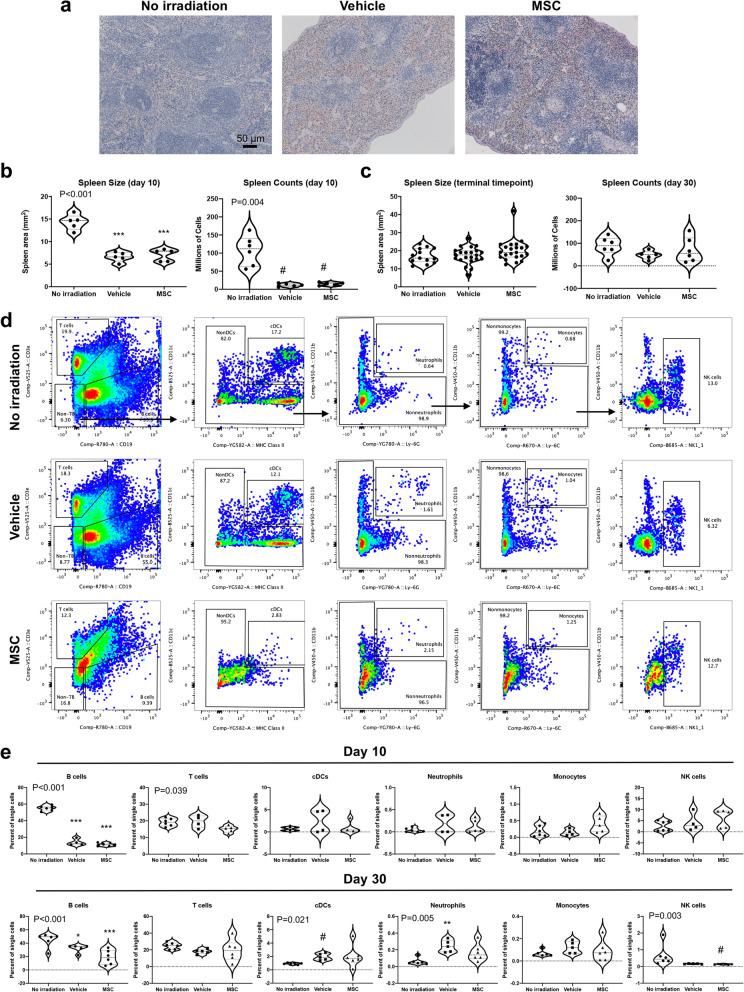
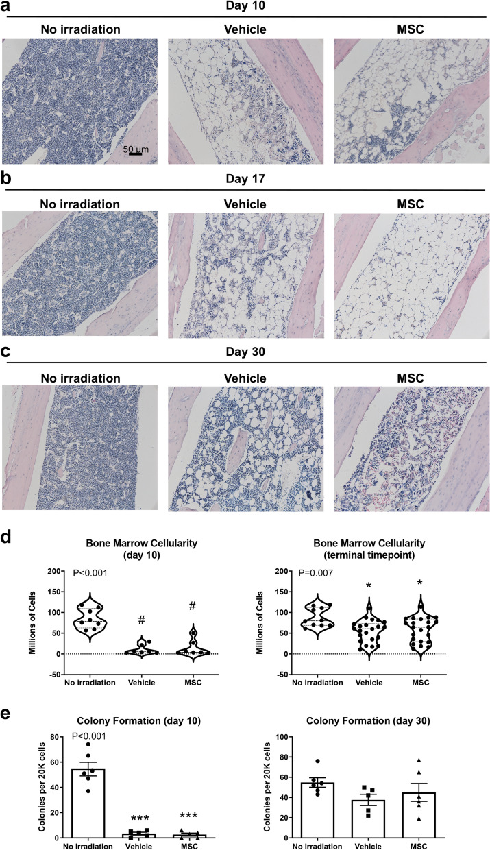
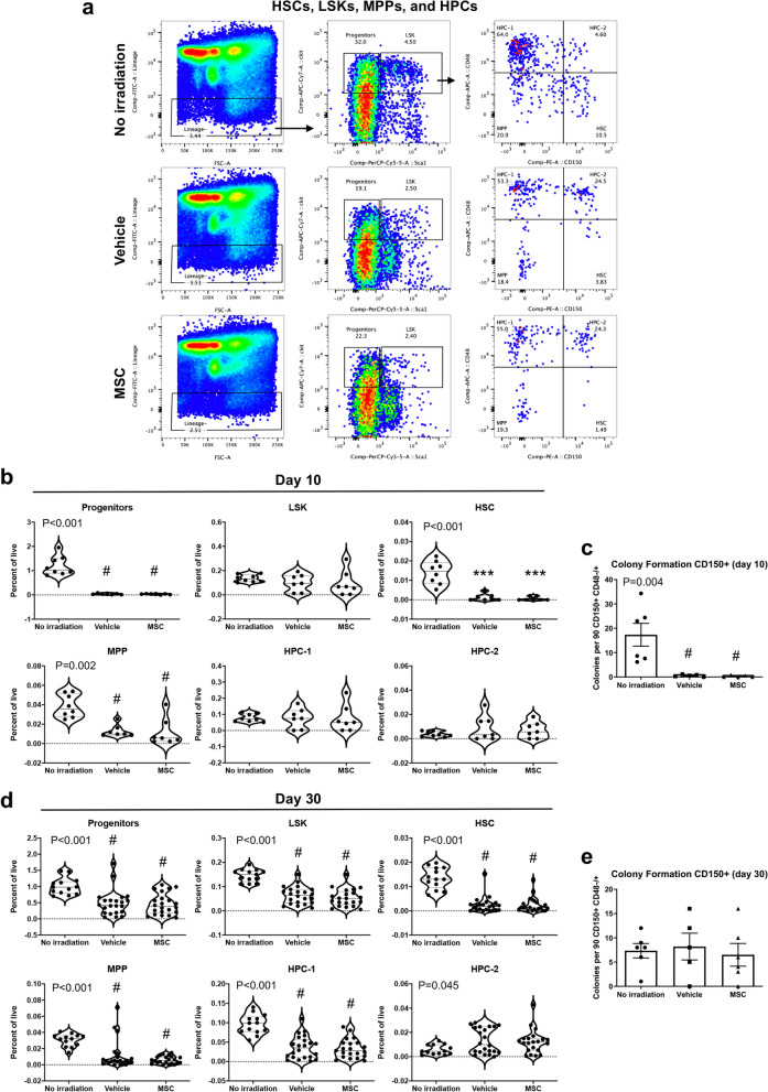
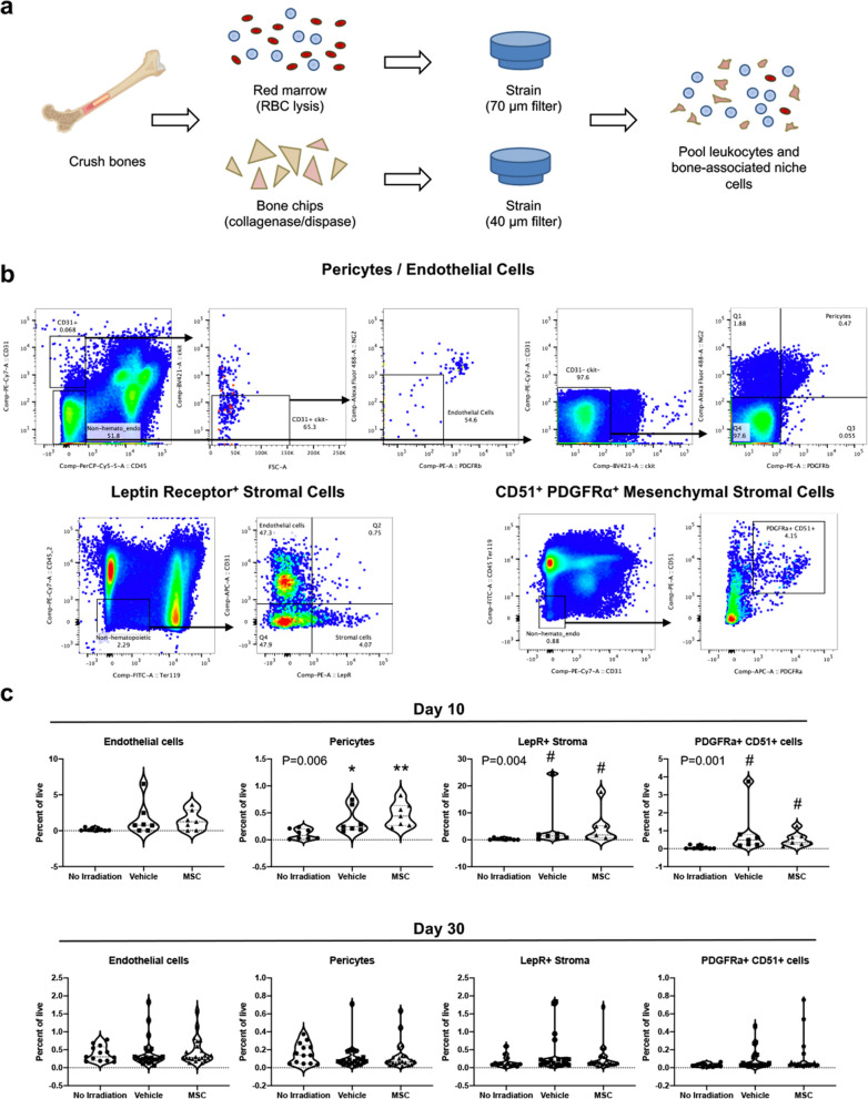
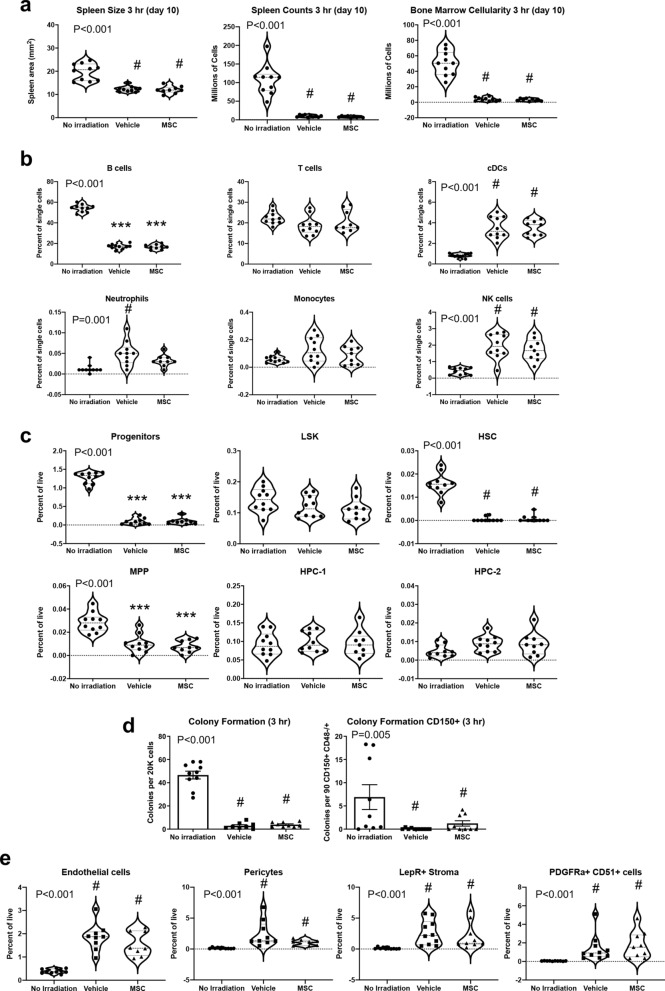
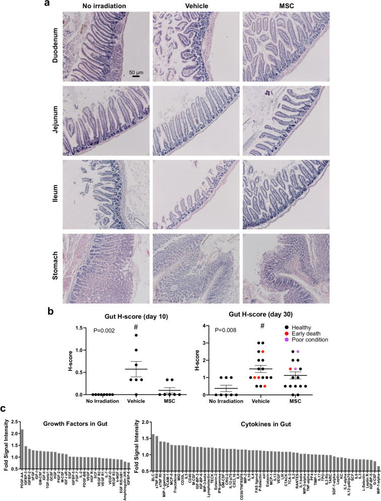
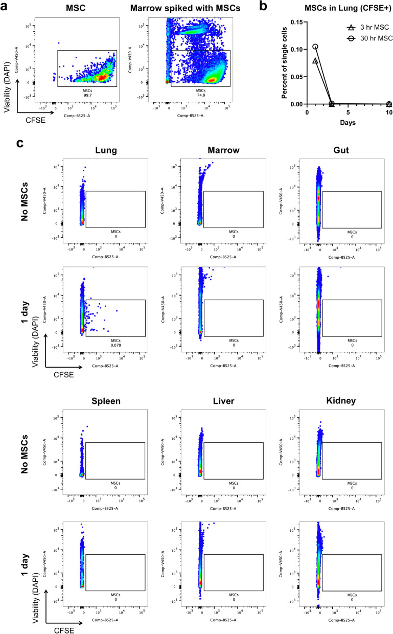
The only available option to treat radiation-induced hematopoietic syndrome is allogeneic hematopoietic cell transplantation, a therapy unavailable to many patients undergoing treatment for malignancy, which would also be infeasible in a radiological disaster. Stromal cells serve as critical components of the hematopoietic stem cell niche and are thought to protect hematopoietic cells under stress. Prior studies that have transplanted mesenchymal stromal cells (MSCs) without co-administration of a hematopoietic graft have shown underwhelming rescue of endogenous hematopoiesis and have delivered the cells within 24 h of radiation exposure. Herein, we examine the efficacy of a human bone marrow-derived MSC therapy delivered at 3 h or 30 h in ameliorating radiation-induced hematopoietic syndrome and show that pancytopenia persists despite MSC therapy. Animals exposed to radiation had poorer survival and experienced loss of leukocytes, platelets, and red blood cells. Importantly, mice that received a therapeutic dose of MSCs were significantly less likely to die but experienced equivalent collapse of the hematopoietic system. The cause of the improved survival was unclear, as complete blood counts, splenic and marrow cellularity, numbers and function of hematopoietic stem and progenitor cells, and frequency of niche cells were not significantly improved by MSC therapy. Moreover, human MSCs were not detected in the bone marrow. MSC therapy reduced crypt dropout in the small intestine and promoted elevated expression of growth factors with established roles in gut development and regeneration, including PDGF-A, IGFBP-3, IGFBP-2, and IGF-1. We conclude that MSC therapy improves survival not through overt hematopoietic rescue but by positive impact on other radiosensitive tissues, such as the intestinal mucosa. Collectively, these data reveal that MSCs could be an effective countermeasure in cancer patients and victims of nuclear accidents but that MSCs alone do not significantly accelerate or contribute to recovery of the blood system.


Conflict of interest statement

B.S.G., C.S.C., and P.L.W. are inventors on a patent for conditioning of stem and progenitor cells for cellular therapy (U.S. Patent US20180187141A1). All other authors declare no conflict of interest.

Figures

Figure 1
Figure 1
Radiation-induced pancytopenia persists despite improvement in survival of MSC recipients. (a) Survival following acute radiation injury is improved with MSC therapy 30 h after exposure (Kaplan–Meier survival plot; Log-rank test, P = 0.0168; n = 26 mice per group). Experiments were conducted across 6 independent experiments on different days. (b) Body weight drops in response to radiation, reaching lowest levels around 17 days (Two-way ANOVA, P = 0.2074 for treatment groups and P < 0.0001 for time; n = 20 mice per group). Experiments were conducted across 4 independent experiments on different days. Body weights are represented as mean ± SEM. (c) White blood cells were depleted within 3 days of radiation and remained low to the endpoint of the study. Platelets and red blood cells experienced a steady drop after 3 days, with nadirs at 17 and 10 days, respectively. Hemoglobin levels drop to a nadir of < 5 g/dL at 17 days after radiation exposure. Elevated mean corpuscular volume corresponds with red blood cell recovery beginning around 21 days after radiation but returns to near normal levels by day 30. Statistical significance of comparisons of treatment group and time by two-way ANOVA are shown on each graph. All data points are plotted for individual animals. WBC white blood cells, RBC red blood cells, Hb hemoglobin, MCV mean corpuscular volume, NE neutrophils, LY lymphocytes, MO monocytes, EO eosinophils, BA basophils.
Figure 2
Figure 2
Radiation injures the spleen and alters immune cell composition. (a) H&E staining of the spleen at day 10 reveals fibrosis and visible hemosiderin, indicative of breakdown of red blood cells, in the irradiated mice regardless of whether they received vehicle or MSCs. (b) Splenic cellularity and size 10 days after irradiation are reduced but are indistinguishable in irradiated and unirradiated mice (c) at the terminal time point. (d) Representative flow cytometry plots of gating for immune cell lineages, including B and T lymphocytes, dendritic cells (cDCs), neutrophils, monocytes, and NK cells. (e) Quantification of splenic immune cell composition at day 10 and day 30. Statistical significance of differences between groups is depicted on the graphs. Posthoc comparisons are demarcated by asterisks *P < 0.05, **P < 0.01, and ***P < 0.001 for Holm–Sidak (One-way ANOVA) or pound symbol #P < 0.5 for Dunn’s (Kruskal–Wallis One-way ANOVA) analyses relative to No irradiation control.
Figure 3
Figure 3
Bone marrow hypocellularity and architectural disruption is not abrogated by MSC therapy. (a) In the proximal tibia, extensive marrow aplasia and adipocytic expansion recognizable as empty round vacuoles are apparent 10 days after injury by H&E staining. (b) Mice that died between 14 and 17 days post-radiation exhibited severe hypocellularity of the bone marrow. Histopathology of two mice that died at day 17 suggests little evidence of hematologic recovery in the marrow. (c) Marrow is partially restored in surviving mice at day 30. (d) Recovery from aplasia was variable but largely reflected incomplete regeneration of the marrow at the study end point at 30 days. (e) CFU activity of whole bone marrow cells. Statistical significance of differences between groups is depicted on the graphs. Posthoc comparisons are demarcated by asterisks *P < 0.05 and ***P < 0.001 for Holm–Sidak (One-way ANOVA) or pound symbol #P < 0.5 for Dunn’s (Kruskal–Wallis One-way ANOVA) analyses relative to No irradiation control.
Figure 4
Figure 4
MSC therapy fails to promote recovery of hematopoietic stem and progenitor cells. (a) Frequencies of HSCs, LSK, HPC-1, HPC-2, MPP, and progenitors were determined by the depicted gating strategies. (b,d) Frequencies of hematopoietic subsets are equivalent in vehicle and MSC recipients. (c,e) CFU activity of sorted CD150+ cells. Statistical significance of differences between groups is depicted on the graphs. Posthoc comparisons are demarcated by asterisks ***P < 0.001 for Holm–Sidak (One-way ANOVA) or pound symbol #P < 0.5 for Dunn’s (Kruskal–Wallis One-way ANOVA) analyses relative to No irradiation control.
Figure 5
Figure 5
Cellular composition of the niche appears unchanged by MSC therapy. (a) Schematic overview of bone marrow isolation for analysis of hematopoietic and non-hematopoietic cells. (b) Representative flow cytometry plots show gating strategies used to define specified niche cell subsets. (c) Loss of hematopoietic cells from the marrow in irradiated mice results in greater relative frequencies of niche cells, although no significant differences exist between vehicle and MSC groups. Statistical significance of differences between groups is depicted on the graphs. Posthoc comparisons are demarcated by asterisks *P < 0.05 and **P < 0.01 for Holm–Sidak (One-way ANOVA) or pound symbol #P < 0.5 for Dunn’s (Kruskal–Wallis One-way ANOVA) analyses relative to No irradiation control.
Figure 6
Figure 6
Administration of MSC therapy 3 h after radiation produces outcomes similar to 30 h infusion. Measurement of (a) spleen size and cellularity, (b) immune lineages in the spleen, (c) HSPC frequencies, (d) colony formation activity of whole bone marrow or sorted CD150+ cells, and (e) niche cell populations in the bone marrow. Statistical significance of differences between groups is depicted on the graphs. Posthoc comparisons are demarcated by asterisks ***P < 0.001 for Holm–Sidak (One-way ANOVA) or pound symbol #P < 0.5 for Dunn’s (Kruskal–Wallis One-way ANOVA) analyses relative to No irradiation control.
Figure 7
Figure 7
Small intestine exhibits signs of injury. (a) Photomicrographs of the small intestine show pathology of the mucosa. (b) H-scores of histopathological analysis of the gut at day 10 and the terminal time point are consistent with greater injury to the small intestine in vehicle treated mice that is less evident in recipients of MSC therapy (Kruskal–Wallis One Way Analysis of Variance on Ranks, Dunn’s Method #P < 0.05 for No irradiation vs. Vehicle). (c) Cytokines and growth factors in the gut are altered by MSC therapy.
Figure 8
Figure 8
Human MSCs lodge in the lung and are undetectable in other organs. (a) CFSE-labeled human MSCs are detectable by flow cytometry when spiked into unlabeled bone marrow. (b) Lung was the only organ in which CFSE+ MSCs could be detected 1 and 3 days after infusion. This distribution occurred for administration of MSCs at 3 h and 30 h after irradiation. (c) Representative flow plots are depicted for all organs analyzed.

References

    1. Mauch P, et al. Hematopoietic stem cell compartment: acute and late effects of radiation therapy and chemotherapy. Int. J. Radiat. Oncol. Biol. Phys. 1995;31:1319–1339. - PubMed
    1. López M, Martín M. Medical management of the acute radiation syndrome. Rep. Pract. Oncol. Radiother. 2011;16:138–146. - PMC - PubMed
    1. Kroemeke A, Sobczyk-Kruszelnicka M, Kwissa-Gajewska Z. Everyday life following hematopoietic stem cell transplantation: decline in physical symptoms within the first month and change-related predictors. Qual. Life Res. 2018;27:125–135. - PMC - PubMed
    1. DiCarlo AL, et al. Cellular therapies for treatment of radiation injury: report from a NIH/NIAID and IRSN workshop. Radiat. Res. 2017;188:e54–e75. - PMC - PubMed
    1. Rios C, Jourdain J-R, DiCarlo AL. Cellular therapies for treatment of radiation injury after a mass casualty incident. Radiat. Res. 2017;188:242–245. - PMC - PubMed

Publication types

MeSH terms