Review
. 2021 Jun;28(6):1804-1821.
doi: 10.1038/s41418-020-00705-8. Epub 2020 Dec 17.

ER-mitochondria contact sites in neurodegeneration: genetic screening approaches to investigate novel disease mechanisms

Affiliations
Review

ER-mitochondria contact sites in neurodegeneration: genetic screening approaches to investigate novel disease mechanisms

Emma Louise Wilson et al. Cell Death Differ. 2021 Jun.

Erratum in

Abstract

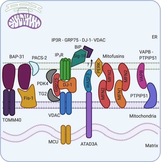
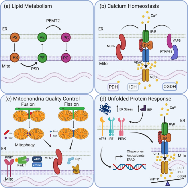
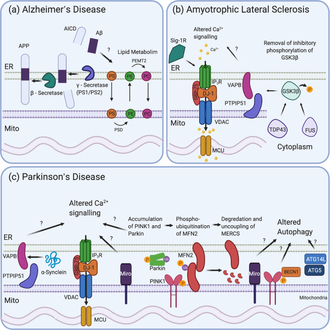
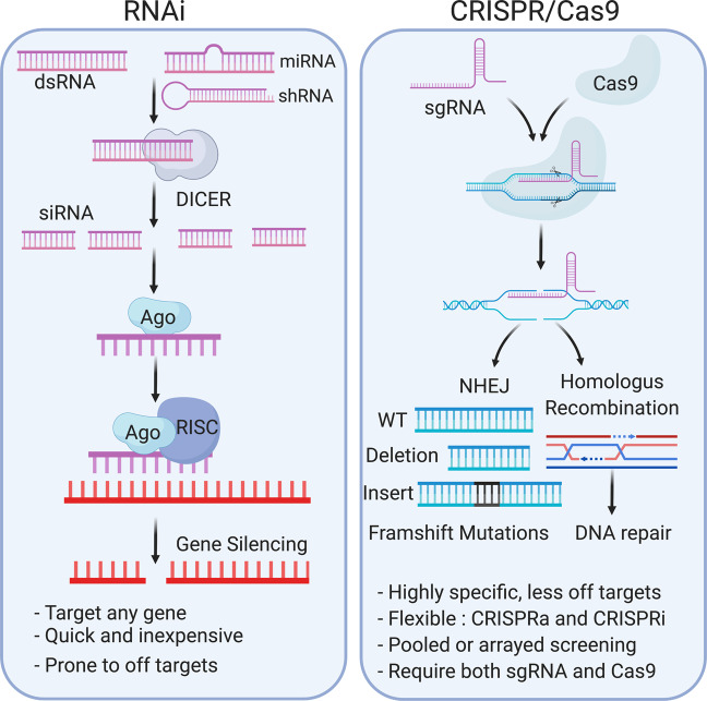
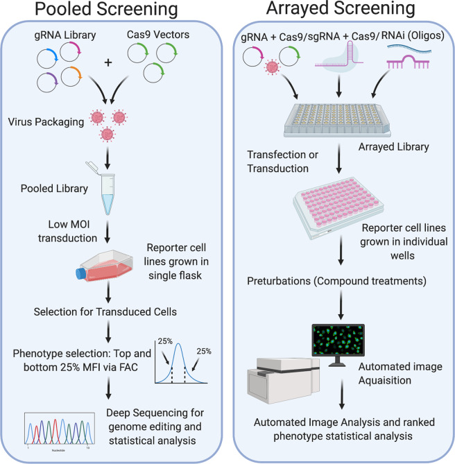
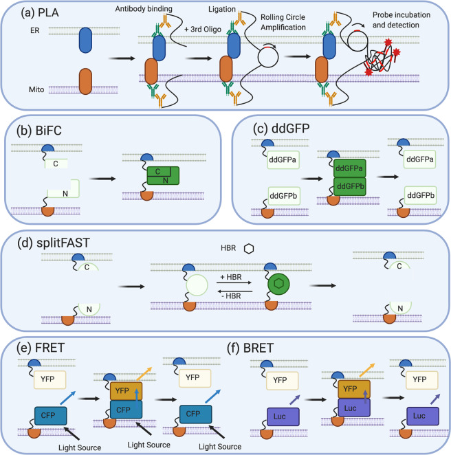
Mitochondria-ER contact sites (MERCS) are known to underpin many important cellular homoeostatic functions, including mitochondrial quality control, lipid metabolism, calcium homoeostasis, the unfolded protein response and ER stress. These functions are known to be dysregulated in neurodegenerative diseases, including Parkinson's disease (PD), Alzheimer's disease (AD) and amyloid lateral sclerosis (ALS), and the number of disease-related proteins and genes being associated with MERCS is increasing. However, many details regarding MERCS and their role in neurodegenerative diseases remain unknown. In this review, we aim to summarise the current knowledge regarding the structure and function of MERCS, and to update the field on current research in PD, AD and ALS. Furthermore, we will evaluate high-throughput screening techniques, including RNAi vs CRISPR/Cas9, pooled vs arrayed formats and how these could be combined with current techniques to visualise MERCS. We will consider the advantages and disadvantages of each technique and how it can be utilised to uncover novel protein pathways involved in MERCS dysfunction in neurodegenerative diseases.


Conflict of interest statement

The authors declare that they have no conflict of interest.

Figures

Fig. 1
Fig. 1. The molecular composition of mitochondria-ER contact sites: several sets of complexes tether the mitochondria and ER.
BAP-31 in the ER interacts with Fis-1 and TOMM40 in the OMM. PACS-2, a multifunctional sorting protein in the ER, regulates BAP-31 MERCS interactions. IP3R in the ER and VDAC in the OMM form a tetramer complex with regulatory proteins, GRP75 and DJ-1, to control calcium (Ca2+) transfer into the mitochondria. Further regulatory proteins such as TG2 and PDK-4 also bind GRP75–IP3R–VDAC complex regulating MERCS. Sig-1R accumulates in MERCS and can stabilise IP3R in MERCS, but also interacts with chaperone protein BIP in the ER lumen. ATAD3A can cross the IMM and OMM to interact with BiP in the ER via the cytosolic protein WASF3 and other unknown proteins. MFN2 is located in both the ER and mitochondrial membrane and can homodimerise with itself or heterodimerise with MFN1 in the OMM. MFN2 has known interactions with PERK, which are required for progression through the UPR. Finally, VAPB in the ER membrane and PTPIP51 in the OMM interact and directly regulate MERCS size and length and act as a physical tether.
Fig. 2
Fig. 2. Cellular functions of mitochondria-ER contact sites.
a Lipid metabolism requires the transfer of phospholipids from the ER to mitochondria and back again at MERCS. Phosphatidylserine (PS) in the ER is transferred to mitochondria where it is converted to phosphatidylethanolamine (PE) by enzyme PS Decarboxylase (PSD). PE is shuttled back to ER where PE-N-methyltransferase (PEMT) modifies it to phosphatidylcholine (PC). b Ca2+ homoeostasis underpins many MERCS functions and is essential to maintain cellular health. The tetramer complex IP3R, VDAC, GRP75 and DJ-1 allow transfer of Ca2+ from ER through IP3R and VDAC to the inter mitochondria membrane space, producing Ca2+ hotspots. These hotspots activate MCU, allowing Ca2+ into the mitochondria matrix that promotes enzymes involved in ATP production, such as PHD, IDH and OGDH. c MERCS impacts on MQC pathways. Both mitochondria fission and fusion require the ER and occur at MERCS. Mitochondrial fusion allows the mixing of damaged and healthy mitochondria components, diluting the damage and helping to maintain the overall health of the mitochondria. Mitochondrial fission can protect the mitochondria network by segregating highly damaged sections, promoting mitophagy. Mitochondrial fission genes Drp1 and Fis-1 and mitochondrial fusion genes, MFN1/2 and OPA1 regulate changes in mitochondrial morphology. MFN2 is found in MERCS, and it is established that the ER constricts the mitochondria, allowing oligomerisation of Drp1 around the mitochondria. MERCS is also involved in mitophagy; both PINK1 and Parkin are found in MERCS under mitophagy induction, as well as key autophagy components ATG5 and ATG14L. d The accumulation of unfolded proteins in the ER increases ER stress and upregulates the chaperone protein BIP that initiates the UPR. BiP activates three pathways of the UPR: ATF6, IRE3 and PERK. Activation of these receptors activates chaperone proteins, antioxidant response proteins and ER-associated protein degradation (ERAD) machinery. ER stress can increase Ca2+ import into the mitochondria to increase the efficiency of Ca2+-dependent enzymes PDH, IDH and IGDH required in ATP production, providing energy for the chaperone machinery. A balance is required, however, as high Ca2+ influx into mitochondria, caused by severe ER stress, can trigger mitochondria permeability transition pore (mPTP) opening, leading to mitochondrial swelling and the initiation of apoptosis.
Fig. 3
Fig. 3. Mitochondrial ER contact sites in neurodegeneration.
a AD: amyloid precursor protein (APP), along with its metabolites, and β- and γ-secretase enzymes are found in MERCS. The APP is first cleaved by β-secretase and then γ-secretase to release Aβ. Aβ has been shown to alter lipid metabolism at MERCS. b ALS: TDP-43 and FUS in the cytoplasm activate GSK3β by dephosphorylating it at cysteine 9. GSK3β can then disrupt VAPB and PTPIP51 binding, uncoupling the ER from the mitochondria and altering Ca2+ signalling. Sig-1R can bind IP3R, stabilising it in the membrane, while loss of Sig-1R can result in ALS-like symptoms in mice and uncoupling of MERCS. c PD: WT or mutated α-Synuclein interacts with VAPB altering its binding to PTPIP51, disrupting MERCS and Ca2+ signalling. Miro is present in MERCS and can disrupt Ca2+ signalling and autophagy. PINK1 and Parkin, under mitophagy induction, have been shown to localise to MERCS and also impact on Ca2+ signalling and to promote the phosphoubiquitination of MFN2, resulting in its degradation and the uncoupling of the mitochondria from the ER. PINK1 and Miro have also been associated with altered mitophagy as PINK1 interacts with BECN1. Key autophagy genes (e.g., ATG14L and ATG5) are also present in MERCS and impact on mitophagy, a pathway dysregulated in PD.
Fig. 4
Fig. 4. Mechanisms for RNAi and CRISPR/Cas9 gene silencing techniques.
In RNAi knockdown, the presence of double-stranded RNA (dsRNA), microRNA (miRNA) and short-hairpin RNA (shRNA) in the cell initiates recruitment of the ribonuclease DICER. DICER cuts dsRNA into shorter fragments called small-interfering RNA (siRNA). Argonaut is a key component of the RNA-induced silencing complex (RISC) that can bind non-coding RNAs, including siRNAs, and recruit the remaining RISC complex components. The RISC complex identifies mRNA of interest, via complimentary base pairing, and cleaves it, inhibiting translation of that protein resulting in a knockdown. The CRISPR/Cas9 system utilises the adaptive immune response of bacteria to conduct genome editing. Both Cas9 endonuclease, as a vector or protein, and gRNAs are required for this process. gRNAs (aka sgRNAs) are short synthetic RNA sequences composed of a tracrRNA, a scaffold sequence necessary to bind Cas9, and crisprRNA (crRNA) (a user-defined 20-bp nucleotide sequence complementary to the gene of interest (GOI)). Through complementary base pairing of the crRNA, the Cas9 is directed to specific genomic locations where it creates double-stranded breaks (DSB). Two principal mechanisms repair DSB: non-homologous end joining (NHEJ) and homologous recombination (HR). NHEJ is error prone and can result in insertions and/or deletions (INDELS) in the genome, causing frameshift mutations and leading to premature stop codons and gene knockout. The HR pathway uses template DNA and DNA synthesis machinery to repair the DNA without error, and can be utilised to incorporate point mutations or other genes (knock-ins).
Fig. 5
Fig. 5. Workflow for pooled and arrayed screening approaches.
Pooled screening requires the gRNA library and Cas9 to be delivered into cells within a single vessel (using a low MOI viral transduction to maintain a 1:1 ratio of gRNA to cells). The cells then undergo selection for transduced cells and further selection for the phenotype of interest. For example, mean fluorescence intensity (MFI) where the top and bottom 25% are examined for survival. The output for a pooled screen comes from deep sequencing of the genomic DNA from control vs treated samples. Statistical analysis of gRNAs that are enriched or depleted is conducted. Arrayed screens can be conducted with a wider range of silencing techniques, including CRISPR/Cas9 or RNAi. This is because the final readout comes from phenotypic analysis, rather than deep sequencing of genomic DNA. Individual gRNAs and siRNAs are delivered (by transduction or transfection) to cells in specific wells of a 96-well plate (eliminating the possibility that more than one gRNA or si9RNA could be delivered per cell). The transfected cells remain in an arrayed format for phenotype analysis, and statistical analysis in which phenotypes of interest can be matched with gRNA/siRNA by their position in the plate.
Fig. 6
Fig. 6. Fluorescence-based techniques to visualise MERCS.
a PLA: the sample is fixed and incubated with primary antibodies targeting proteins in MERCS (usually MERCS tethers) and a modified secondary antibody (which is attached to an oligonucleotide strand). If the two proteins are in close proximity, as they are in MERCS, the addition of a third oligonucleotide results in complementary base pairing between the three oligonucleotides and the formation of circular DNA. The circular DNA can undergo rolling circle amplification, creating a long strand of DNA and multiple copies of the circular DNA. Fluorescent probes are designed to be complementary to a sequencing within the circular DNA. The probes hybridise in multiple regions, allowing the visualisation of the fluorescent signal and MERCS. b BiFC also utilises proximity to visualise MERCS. Two non-fluorescence fragments of a fluorescence protein, commonly GFP or Venus, are fused to transmembrane domains, or whole proteins, found in the ER or mitochondrial membrane. If MERCS does not form the BiFc, constructs remain separate and do not fluoresce, but when MERCS forms, the ER and mitochondria membrane come into close proximity bringing with them the two non-fluorescent fragments, allowing them refold into whole GFP or Venus protein whose fluorescence can be visualised and measured. c Similarly, the ddGFP system is fused to protein fragments or whole proteins found in ER and mitochondria membrane; however, each protein is fused to non-fluorescence ddGFP monomers. One monomer contains a chromophore that is destabilised and quenched, while the other completely lacks a chromophore. When MERCS forms, these ddGFP monomers come into close proximity, heterodimerise and complement each other, allowing fluorescent detection. As there is no protein folding, this process is reversible. d splitFAST utilises the 14- kda protein (FAST), which is split into N- and C-terminal fragments. These fragments can be attached to two interacting proteins or membrane fragments of a protein. For the study of MERCS, these interacting proteins would be in the ER or mitochondrial membrane. When MERCS forms, the N and C fragments combine, and upon the addition of HBR, they fluoresce allowing visualisation of MERCS. This is reversible, as HBR can be added or removed, but the FAST protein can also dissociate when the ER and mitochondria membrane are not in close proximity. e FRET: a FRET donor (CFP) and FRET acceptor (YFP) are fused to resident ER or mitochondrial proteins or transmembrane protein fragments. When ER or mitochondrial membranes are not in close proximity, a light source illuminates the CFP donor but the YFP acceptor is not close enough for FRET to occur and so blue light emitted. When MERCS forms, the FRET donor (CFP) and acceptor (YFP) are in close proximity, so FRET can occur, and blue light is transferred to YFP and yellow fluorescence is emitted. This is reversible as no protein folding or contact occurs between FRET pairs, f BRET also employs FRET to visualise MERCS; however, rather than CFP, the resident ER, mitochondria protein or transmembrane fragments are fused to a luciferase enzyme that acts as a light source. When MERCS forms, the luciferase enzyme and FRET acceptor are in close proximity, and so energy transfer can occur from the luciferase enzyme and YFP, which is emitted as yellow florescence. This is reversible as no protein folding occurs.

References

    1. Bernhard W, Rouiller C. Close topographical relationship between mitochondria and ergastoplasm of liver cells in a definite phase of cellular activity. J Biophys Biochem Cytol. 1956. 10.1083/jcb.2.4.73. - PMC - PubMed
    1. Giacomello M, Pellegrini L. The coming of age of the mitochondria-ER contact: a matter of thickness. Cell Death Differ. 2016. 10.1038/cdd.2016.52. - PMC - PubMed
    1. Paillusson S, Stoica R, Gomez-Suaga P, Lau DHW, Mueller S, Miller T, et al. There’s Something Wrong with my MAM; the ER-Mitochondria Axis and Neurodegenerative Diseases. Trends Neurosci. 2016. 10.1016/j.tins.2016.01.008. - PMC - PubMed
    1. Csordás G, Renken C, Várnai P, Walter L, Weaver D, Buttle KF, et al. Structural and functional features and significance of the physical linkage between ER and mitochondria. J Cell Biol. 2006;174:915–21. doi: 10.1083/jcb.200604016. - DOI - PMC - PubMed
    1. Basso V, Marchesan E, Ziviani E. A trio has turned into a quartet: DJ-1 interacts with the IP3R-Grp75-VDAC complex to control ER-mitochondria interaction. Cell Calcium. 2020. 10.1016/j.ceca.2020.102186. - PubMed

Publication types